

β‑Ecdysterone promotes autophagy and inhibits apoptosis in osteoporotic rats
- PMID: 29138818
- PMCID: PMC5780099
- DOI: 10.3892/mmr.2017.8053
β‑Ecdysterone promotes autophagy and inhibits apoptosis in osteoporotic rats
Abstract
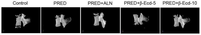
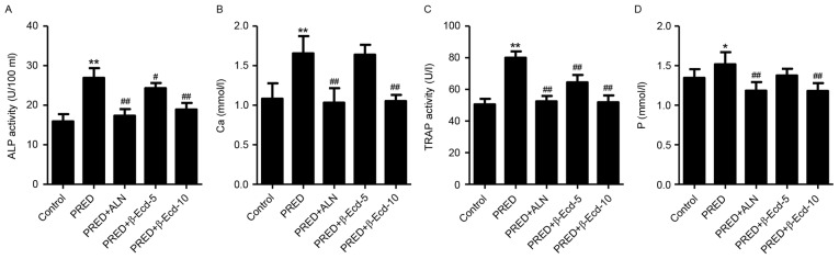
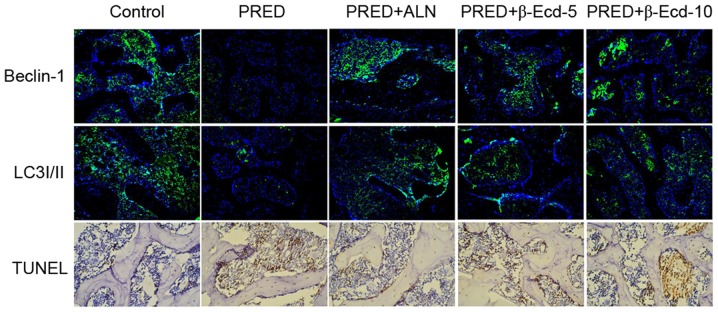
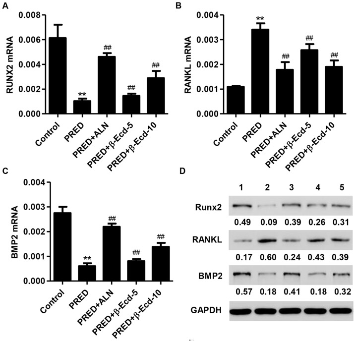
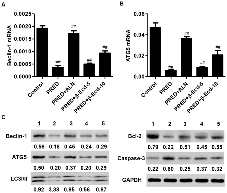
Osteoporosis is an aging process of skeletal tissues with characteristics of reductions in bone mass and microarchitectural deterioration of bone tissue. The present study aimed to investigate the effects of glucocorticoid‑induced osteoporosis on osteoblasts and to examine the roles of β‑ecdysterone (β‑Ecd) involved. In the present study, an in vivo model of osteoporosis was established through the subcutaneous implantation of prednisolone (PRED) into Sprague‑Dawley rats, with or without a subcutaneous injection of β‑Ecd (5 or 10 mg/kg body weight). Expression of Beclin‑1 and microtubule‑associated protein 1A/1B‑light chain 3I/II and apoptosis in lumbar vertebrae tissues was measured by immunofluorescence and TUNEL assays, respectively. Serum concentration of calcium and phosphorus, and the activity of tartrate‑resistant acid phosphatase (TRAP) and alkaline phosphatase (ALP) were measured by biochemical assay. Reverse transcription‑quantitative polymerase chain reaction and western blotting was used for detect the expression of related genes and proteins. PRED treatment inhibited bone formation by decreasing bone mineral density, and suppressing the expression of Runt‑related transcription factor 2 and bone morphogenetic protein 2, while enhancing the activity of alkaline phosphatase, upregulating the expression of receptor activator of nuclear factor-κB ligand, and increasing the serum content of calcium, phosphorus and tartrate‑resistant acid phosphatase in rats. Additionally, PRED was revealed to inhibit autophagy through the downregulation of Beclin‑1, autophagy protein 5 and microtubule‑associated protein 1A/1B‑light chain 3I/II expression, whereas it induced the apoptosis, through the activation of caspase‑3 and the suppression of apoptosis regulator BCL2 expression. Notably, the PRED‑induced alterations in bone formation, autophagy and apoptosis were revealed to be attenuated by β‑Ecd administration. In conclusion, the findings of the present study suggested that β‑Ecd may be a promising candidate for the development of therapeutic strategies for the treatment of osteoporosis, through the induction of autophagy and the inhibition of apoptosis in vivo.
Figures
Similar articles
-
Effect of β‑ecdysterone on glucocorticoid‑induced apoptosis and autophagy in osteoblasts.Mol Med Rep. 2018 Jan;17(1):158-164. doi: 10.3892/mmr.2017.7840. Epub 2017 Oct 20. Mol Med Rep. 2018. PMID: 29115419 Free PMC article.
-
Fangchinoline Promotes Autophagy and Inhibits Apoptosis in Osteoporotic Rats.Med Sci Monit. 2019 Jan 11;25:324-332. doi: 10.12659/MSM.912624. Med Sci Monit. 2019. Retraction in: Med Sci Monit. 2022 Oct 24;28:e938703. doi: 10.12659/MSM.938703. PMID: 30632520 Free PMC article. Retracted.
-
Rapamycin reduces severity of senile osteoporosis by activating osteocyte autophagy.Osteoporos Int. 2016 Mar;27(3):1093-1101. doi: 10.1007/s00198-015-3325-5. Epub 2015 Sep 22. Osteoporos Int. 2016. PMID: 26395886
-
Autophagy: A Promising Target for Age-related Osteoporosis.Curr Drug Targets. 2019;20(3):354-365. doi: 10.2174/1389450119666180626120852. Curr Drug Targets. 2019. PMID: 29943700 Review.
-
Targeting autophagy in osteoporosis: From pathophysiology to potential therapy.Ageing Res Rev. 2020 Sep;62:101098. doi: 10.1016/j.arr.2020.101098. Epub 2020 Jun 12. Ageing Res Rev. 2020. PMID: 32535273 Review.
Cited by
-
Molecular Targets of 20-Hydroxyecdysone in Mammals, Mechanism of Action: Is It a Calorie Restriction Mimetic and Anti-Aging Compound?Cells. 2025 Mar 13;14(6):431. doi: 10.3390/cells14060431. Cells. 2025. PMID: 40136680 Free PMC article. Review.
-
Ortho-silicic Acid Plays a Protective Role in Glucocorticoid-Induced Osteoporosis via the Akt/Bad Signal Pathway In Vitro and In Vivo.Biol Trace Elem Res. 2023 Feb;201(2):843-855. doi: 10.1007/s12011-022-03201-x. Epub 2022 Mar 21. Biol Trace Elem Res. 2023. PMID: 35314965
-
An Arthropod Hormone, Ecdysterone, Inhibits the Growth of Breast Cancer Cells via Different Mechanisms.Front Pharmacol. 2020 Oct 30;11:561537. doi: 10.3389/fphar.2020.561537. eCollection 2020. Front Pharmacol. 2020. PMID: 33192507 Free PMC article.
-
Knockdown of SMYD3 by RNA Interference Regulates the Expression of Autophagy-Related Proteins and Inhibits Bone Formation in Fluoride-Exposed Osteoblasts.Biol Trace Elem Res. 2025 Apr;203(4):2013-2028. doi: 10.1007/s12011-024-04327-w. Epub 2024 Aug 6. Biol Trace Elem Res. 2025. PMID: 39106008 Free PMC article.
-
Unraveling the potential mechanisms of the anti-osteoporotic effects of the Achyranthes bidentata-Dipsacus asper herb pair: a network pharmacology and experimental study.Front Pharmacol. 2023 Oct 2;14:1242194. doi: 10.3389/fphar.2023.1242194. eCollection 2023. Front Pharmacol. 2023. PMID: 37849727 Free PMC article.
References
-
- Huang Y, Bo Y, Wu X, Wang Q, Qin F, Zhao L, Xiong Z. An intergated serum and urinary metabonomic research based on UPLC-MS and therapeutic effects of Gushudan on prednisolone-induced osteoporosis rats. J Chromatogr B Analyt Technol Biomed Life Sci. 2016;1027:119–130. doi: 10.1016/j.jchromb.2016.05.019. - DOI - PubMed
MeSH terms
Substances
LinkOut - more resources
Full Text Sources
Other Literature Sources
Medical
Research Materials
