Review
. 2018 Jan;17(1):1403-1412.
doi: 10.3892/mmr.2017.8038. Epub 2017 Nov 13.

Role of innate lymphoid cells in obesity and metabolic disease (Review)

Affiliations
Review

Role of innate lymphoid cells in obesity and metabolic disease (Review)

Jirakrit Saetang et al. Mol Med Rep. 2018 Jan.

Abstract

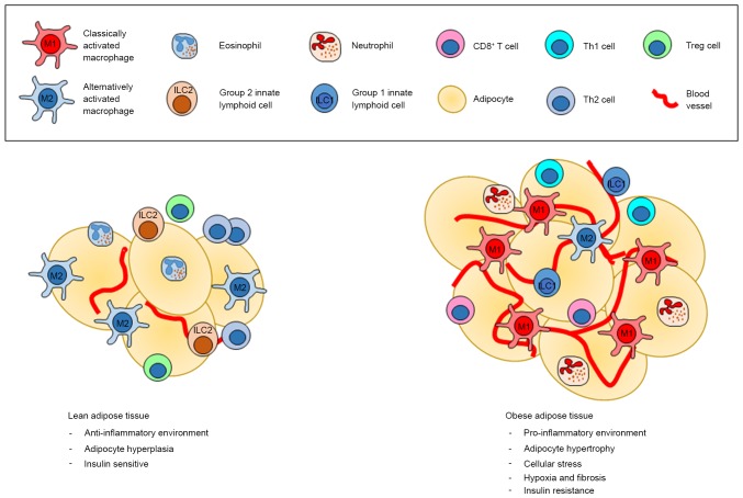
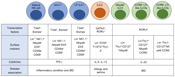
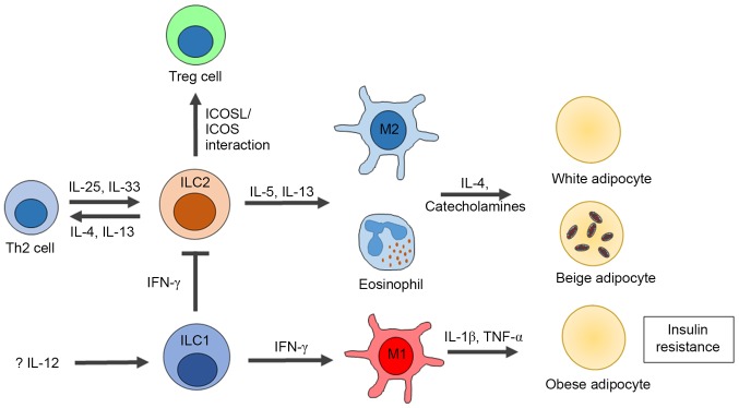
The immune system has previously been demonstrated to be associated with the pathophysiological development of metabolic abnormalities. However, the mechanisms linking immunity to metabolic disease remain to be fully elucidated. It has previously been suggested that innate lymphoid cells (ILCs) may be involved in the progression of numerous types of metabolic diseases as these cells act as suppressors and promoters for obesity and associated conditions, and are particularly involved in adipose tissue inflammation, which is a major feature of metabolic imbalance. Group 2 ILCs (ILC2s) have been revealed as anti‑obese immune regulators by secreting anti‑inflammatory cytokines and promoting the polarization of M2 macrophages, whereas group 1 ILCs (ILC1s), including natural killer cells, may promote adipose tissue inflammation via production of interferon‑γ, which in turn polarizes macrophages toward the M1 type. The majority of studies to date have demonstrated the pathological association between ILCs and obesity in the context of adipose tissue inflammation, whereas the roles of ILCs in other organs which participate in obesity development have not been fully characterized. Therefore, identifying the roles of all types of ILCs as central components mediating obesity‑associated inflammation, is of primary concern, and may lead to the discovery of novel preventative and therapeutic interventions.


Figures

Figure 1.
Figure 1.
The different environment between lean and obese adipose tissues. The most common immune cells found in lean adipose tissue are M2 macrophages, which create the anti-inflammatory environment in cooperation with ILC2s, eosinophils, Treg and Th2 cells. On the other hand, obese adipose tissue is dominated by M1 macrophages, neutrophils, ILC1, Th1 as well as CD8+ T cells, which all promote an inflammatory condition, which in turn support insulin resistance. Treg, regulatory T cells; ILC, innate lymphoid cell.
Figure 2.
Figure 2.
The important transcription factors and cell surface markers for each type of innate lymphoid cells. *Lin = TCRβ+ CD3ε+ CD19+ TCRγδ+ Ly6G+ F4/80+ cells. ILC, innate lymphoid cell; ieILC1, intraepithelial ILC1; LP ILC1, lamina propria-resident ILC1l; ILC1, group 1 ILC; ILC2, group 2 ILC; RORα, retinoic acid-related orphan receptor α; RORγt, retinoic acid receptor-related orphan receptor γt; IBD, inflammatory bowel disease.
Figure 3.
Figure 3.
Influences of ILCs on obesity and insulin resistance. In the healthy state, adipose tissue inflammation is suppressed by IL-5 and IL-13, secreted by ILC2s that contribute to the activation of M2 macrophages and eosinophils. Moreover, the productions of IL-4 and IL-13 from ILC2s also promote Th2 development. The inflammatory condition of adipose tissue in obesity is associated with increased infiltration of M1 macrophages, neutrophils, Th1 and CD8+ T cells. Recently, the role of ILC1 s in adipose tissue inflammation has been identified, which are mediated by the secretion of IFN-γ that promote M1 macrophage polarization and ILC2 suppression. ILC, innate lymphoid cell; ILC1, group 1 ILC; ILC2, group 2 ILC; Treg, regulatory T cells; TNF-α, tumor necrosis factor-α.

Similar articles

Cited by

References

    1. Boulangé CL, Neves AL, Chilloux J, Nicholson JK, Dumas ME. Impact of the gut microbiota on inflammation, obesity and metabolic disease. Genome Med. 2016;8:42. doi: 10.1186/s13073-016-0303-2. - DOI - PMC - PubMed
    1. Lu Y, Loos RJ. Obesity genomics: Assessing the transferability of susceptibility loci across diverse populations. Genome Med. 2013;5:55. doi: 10.1186/gm459. - DOI - PMC - PubMed
    1. Westerterp KR, Plasqui G. Physically active lifestyle does not decrease the risk of fattening. PLoS One. 2009;4:e4745. doi: 10.1371/journal.pone.0004745. - DOI - PMC - PubMed
    1. DiNicolantonio JJ, O'Keefe JH, Lucan SC. Added fructose: A principal driver of type 2 diabetes mellitus and its consequences. Mayo Clin Proc. 2015;90:372–381. doi: 10.1016/j.mayocp.2014.12.019. - DOI - PubMed
    1. DiNicolantonio JJ, Lucan SC, O'Keefe JH. The evidence for saturated fat and for sugar related to coronary heart disease. Prog Cardiovasc Dis. 2016;58:464–472. doi: 10.1016/j.pcad.2015.11.006. - DOI - PMC - PubMed