Comparative Study
. 2018 May;34(5):673-682.
doi: 10.1007/s10554-017-1275-z. Epub 2017 Nov 14.

Vessel centerline reconstruction from non-isocentric and non-orthogonal paired monoplane angiographic images

Affiliations
Comparative Study

Vessel centerline reconstruction from non-isocentric and non-orthogonal paired monoplane angiographic images

Mie Kunio et al. Int J Cardiovasc Imaging. 2018 May.

Abstract

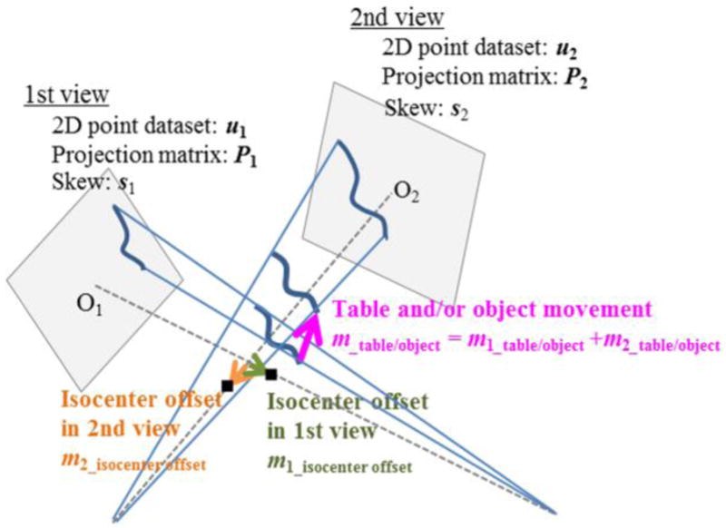
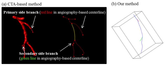
Purpose: Three-dimensional reconstruction of a vessel centerline from paired planar coronary angiographic images is critical to reconstruct the complex three-dimensional structure of the coronary artery lumen and the relative positioning of implanted devices. In this study, a new vessel centerline reconstruction method that can utilize non-isocentric and non-orthogonal pairs of angiographic images was developed and tested.

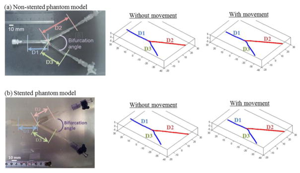
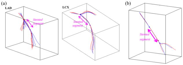
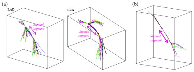
Methods: Our new method was developed in in vitro phantom models of bifurcated coronary artery with and without stent, and then tested in in vivo swine models (twelve coronary arteries). This method was also validated using data from six patients.

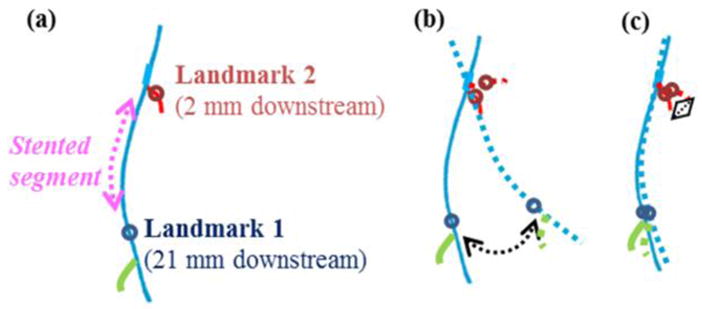
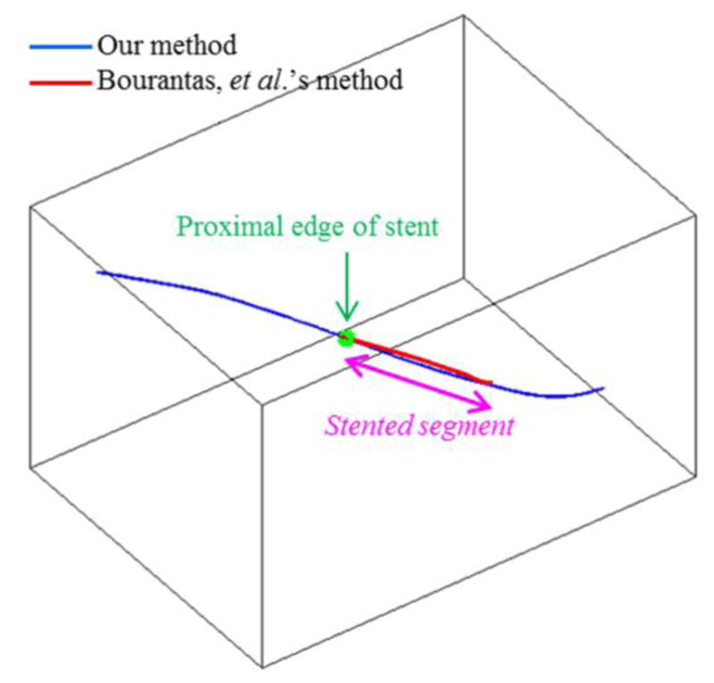
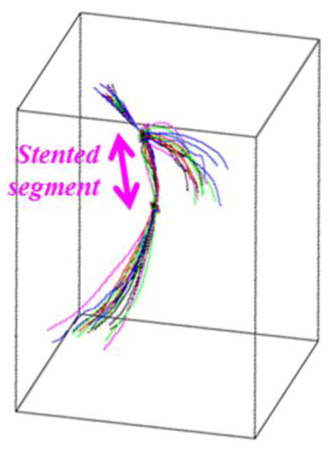
Results: Our new method demonstrated high accuracy (root mean square error = 0.27 mm or 0.76 pixel), and high reproducibility across a broad imaging angle (20°-130°) and between different cardiac cycles in vitro and in vivo. Use of this method demonstrated that the vessel centerline in the stented segment did not deform significantly over a cardiac cycle in vivo. In addition, the total movement of the isocenter in each image could be accurately estimated in vitro and in vivo. The performance of this new method for patient data was similar to that for in vitro phantom models and in vivo animal models.

Conclusions: We developed a vessel centerline reconstruction method for non-isocentric and non-orthogonal angiographic images. It demonstrated high accuracy and good reproducibility in vitro, in vivo, and in clinical setting, suggesting that our new method is clinically applicable despite the small sample size of clinical data.

Keywords: Coronary angiography; Image reconstruction; Reconstruction algorithm; Stereoscopic theory.


Figures

Fig. 1
Fig. 1
Potential causes of the isocenter movement. (a) Machine-origin isocenter offset: The angiography system itself has an isocenter offset when it is rotated. This offset appears in each image as the difference between the center of view and the projected origin. (b) Movement of the object center: During the procedure, the center of the object may move because physicians may move the table and/or patients themselves may move.
Fig. 2
Fig. 2
Algorithm for vessel centerline registration. First, two landmarks, Landmark 1 and 2 were set (a). Then, the secondary geometry (dotted line) was rotated about the origin until the distance between corresponding Landmarks 1 was minimized (b). Next, a vector through the origin and Landmark 1 was defined. Finally, the secondary geometry was rotated again about this vector until the distance between corresponding Landmarks 2 was minimized (c).
Fig. 3
Fig. 3
Phantom models and reconstructed centerline geometries. All the centerlines reconstructed from various imaging angle differences (20°–130°) were similar in lengths and bifurcation angle to the actual geometry in both non-stented and stented phantom models.
Fig. 4
Fig. 4
Comparison between our vessel centerline reconstruction method and the state-of-the-art method. The vessel centerline reconstructed with our method had a quite similar shape to that obtained with Bourantas et al.’s method in the stented segment.
Fig. 5
Fig. 5
Reproducibility across various imaging angle differences (20°–130°). All the vessel centerlines from non-orthogonal pairs of angiographic images (20°–130°) were aligned well to the centerline from an orthogonal pair of angiographic images.
Fig. 6
Fig. 6
Reconstructed vessel centerlines from two different cardiac cycles: (A) pre-clinical results, (B) one patient result. The vessel centerlines that were reconstructed from two different cardiac cycles had quite similar shapes, especially in the stented segment, in both pre-clinical and clinical settings. Blue and red lines represent the vessel centerlines from two different cardiac cycles.
Fig. 7
Fig. 7
Deformation of the vessel centerline over a cardiac cycle: (A) pre-clinical results, (B) one patient result. The vessel centerline did not deform significantly within the stented segment in both pre-clinical and clinical settings. Blue, red, green, and black lines represent the vessel centerlines at end-diastolic, mid-systolic, end-systolic, and mid-diastolic phases, respectively.
Fig. 8
Fig. 8
Reconstructed vessel centerlines with CTA-based method (a) and with our method (b) (one patient result).

Similar articles

Cited by

References

    1. Brown BG, Bolson E, Frimer M, Dodge HT. Quantitative coronary arteriography: estimation of dimensions, hemodynamic resistance, and atheroma mass of coronary artery lesions using the arteriogram and digital computation. Circulation. 1977;55(2):329–337. doi: 10.1161/01.cir.55.2.329. - DOI - PubMed
    1. Thompson CA. Textbook of Cardiovascular Intervention. 1. Springer; 2014. - DOI
    1. Dowe DA, Fioranelli M, Pavone P. Imaging Croronary Arteries. 2. Springer; 2013.
    1. Chen SY, Carroll Jd, Messenger JC, Messenger JC. Quantitative analysis of reconstructed 3-D coronary arterial tree and intracoronary devices. IEEE Trans Med Imaging. 2002;21(7):724–749. - PubMed
    1. Dvir D, Marom H, Guetta V, Kornowski R. Three-dimensional coronary reconstruction from routine single-plane coronary angiograms: in vivo quantitative validation. Int J Cardiovasc Intervent. 2005;7:141–145. - PubMed

MeSH terms