Review
. 2018 Feb;191(2):133-148.
doi: 10.1111/cei.13081. Epub 2017 Dec 27.

Immune checkpoint inhibitors: new strategies to checkmate cancer

Affiliations
Review

Immune checkpoint inhibitors: new strategies to checkmate cancer

R A M Wilson et al. Clin Exp Immunol. 2018 Feb.

Abstract

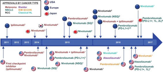
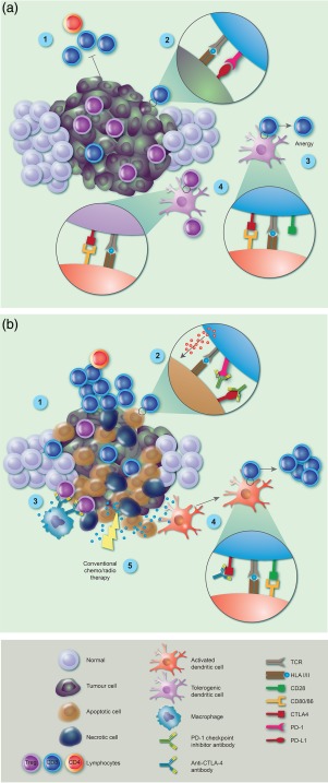
Immune checkpoint inhibitors (ICIs) targeting cytotoxic T lymphocyte-associated protein-4 (CTLA-4) or programmed cell death protein 1 (PD-1) receptors have demonstrated remarkable efficacy in subsets of patients with malignant disease. This emerging treatment modality holds great promise for future cancer treatment and has engaged pharmaceutical research interests in tumour immunology. While ICIs can induce rapid and durable responses in some patients, identifying predictive factors for effective clinical responses has proved challenging. This review summarizes the mechanisms of action of ICIs and outlines important preclinical work that contributed to their development. We explore clinical data that has led to disease-specific drug licensing, and highlight key clinical trials that have revealed ICI efficacy across a range of malignancies. We describe how ICIs have been used as part of combination therapies, and explore their future prospects in this area. We conclude by discussing the incorporation of these new immunotherapeutics into precision approaches to cancer therapy.

Keywords: T cell; antibodies; cancer; tumour immunology.


Figures

Figure 1
Figure 1
History of checkpoint inhibitors: key milestones. Timeline showing when checkpoint inhibitors were approved for the treatment of specific cancers in the United States, Europe and Japan. Tumour type is indicated by the colour of each immune checkpoint inhibitor (ICI) depicted in the figure, according to the key in the top left. Abbreviations: 1L = first‐line; 2L = second‐line; NSCLC = non‐small‐cell lung cancer; NSQ = non‐squamous; PD‐L1 = programmed death ligand 1; RCC = renal cell carcinoma; R/M = recurrent/metastatic; SCCHN = squamous cell carcinoma of the head and neck; SQ = squamous. 1 US Food and Drug Administration. http://www.fda.gov, accessed 11 November 2016. 2 European Medicines Agency. http://www.ema.europa.eu, accessed 11 November 2016. 3ONO Pharmaceutical Co. Ltd [press release], 4 July 2014. 4ONO Pharmaceutical Co. Ltd [press release]. March 23, 2015. 5 ONO Pharmaceutical Co, Ltd. [press release], 17 December 17, 2015. 6Merck [press release], 27 June 2016, accessed 8 August 2016. 7Bristol‐Myers Squibb Company [press release], 22 November 2016. 8Merck [press release], 10 December 2016.
Figure 2
Figure 2
Immune checkpoint inhibitor (ICI) blockade reverses tumour‐mediated immune suppression. (a) Established tumours block immune attack through a variety of mechanisms, including inhibition of tumour‐specific cytotoxic T lymphocyte (CTL) and CD4 T cell activation and function (1). This is driven by tumour over‐expression of programmed cell death protein 1(PD‐L1), interacting with tumour‐specific T cell PD‐1 receptor (2) and T cell anergy induced by tumour‐mediated T cell expression of cytotoxic T lymphocyte‐associated protein 4 (CTLA‐4) inhibitory receptor (4). In addition, tolerogenic dendritic cells (DC) drive regulatory T cell (Treg) induction and expansion via CTLA‐4 (3) and accumulation of Tregs then contributes to the immunosuppressive milieu of the tumour microenvironment (TME). (b) After ICI therapy, there is re‐activation and proliferation of tumour‐specific CTLs via blockade of the PD‐1 axis (1), and return of functional cytotoxicity, resulting in perforin release and tumour cell killing (2). As tumour damage increases, the TME is disrupted allowing macrophages to deplete Tregs via fragment crystalline receptor (FcR) binding of anti‐CTLA‐4 antibody (3). Tumour antigen release is driven by immune lysis of tumour cells which are processed by conventional DC and presented to naive T cells in context of checkpoint inhibition of CTLA‐4, enhancing CTL proliferation and function (4). Tumour damage and antigen release is also supplemented by concomitant use of conventional chemo/radiotherapy, which can reveal new tumour‐associated antigens and contribute to anti‐tumour immune responses (5).

References

    1. Gajewski TF, Schreiber H, Fu YX. Innate and adaptive immune cells in the tumor microenvironment. Nat Immunol 2013; 14:1014–22. - PMC - PubMed
    1. Speiser DE, Ho PC, Verdeil G. Regulatory circuits of T cell function in cancer. Nat Rev Immunol 2016; 16:599–611. - PubMed
    1. Dunn GP, Old LJ, Schreiber RD. The immunobiology of cancer immunosurveillance and immunoediting. Immunity 2004; 21:137–48. - PubMed
    1. Prendergast GC. Immune escape as a fundamental trait of cancer: focus on IDO. Oncogene 2008; 27:3889–900. - PubMed
    1. Igney FH, Krammer PH. Immune escape of tumors: apoptosis resistance and tumor counterattack. J Leukoc Biol 2002; 71:907–20. - PubMed

MeSH terms