. 2017 Nov;33(S1):S81-S92.
doi: 10.1089/aid.2017.0160.

IL-21 Therapy Controls Immune Activation and Maintains Antiviral CD8+ T Cell Responses in Acute Simian Immunodeficiency Virus Infection

Affiliations

IL-21 Therapy Controls Immune Activation and Maintains Antiviral CD8+ T Cell Responses in Acute Simian Immunodeficiency Virus Infection

Gema Méndez-Lagares et al. AIDS Res Hum Retroviruses. 2017 Nov.

Abstract

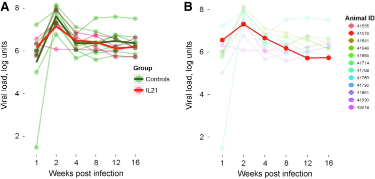
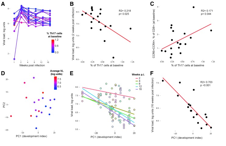
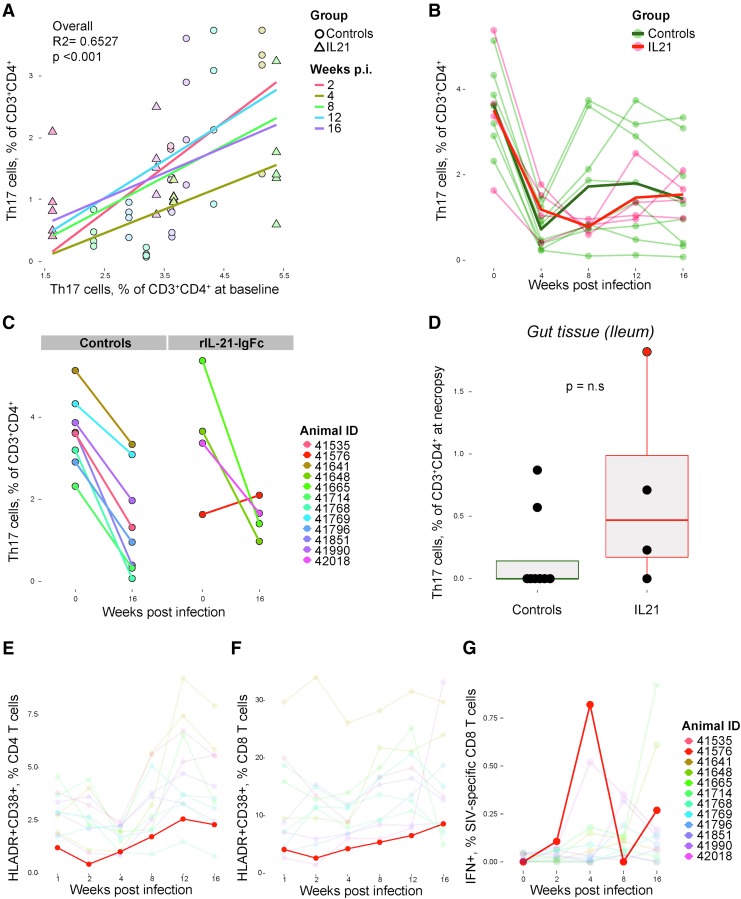
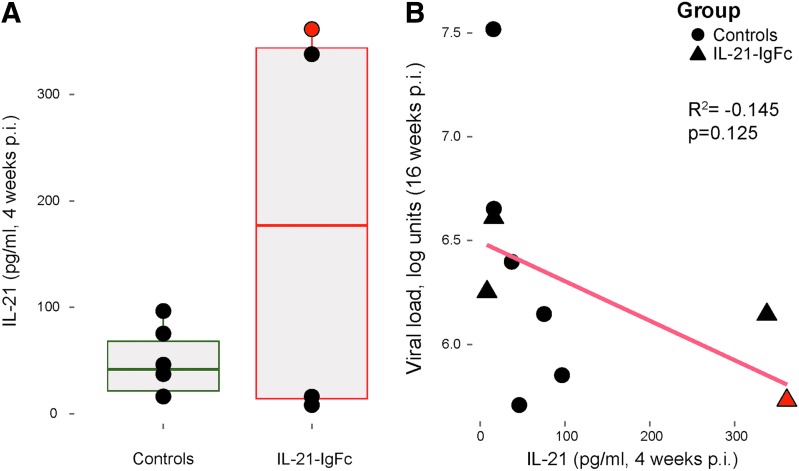
Human immunodeficiency virus (HIV) and simian immunodeficiency virus (SIV) replicate during acute infection in lymphocytes of the gastrointestinal tract, before disseminating systemically. Localized replication and associated loss of gut-resident CD4+ T cells occur regardless of the portal of entry of the virus (e.g., intravenous vs. rectal). Thus, HIV and SIV are tropic for gut tissue, and their pathogenesis requires the special environment of the intestine. T helper 17 (Th17) cells are important contributors to microbial defense in the gut that are vulnerable to HIV infection and whose loss is associated with translocation of microbial products to the systemic circulation, leading to chronic immune activation and disease progression. Interleukin (IL)-21 promotes differentiation and survival of Th17 cells and stimulates CD8+ T cell function. By promoting Th17 cell survival, IL-21 could limit bacterial translocation and immune activation in the setting of acute or rebounding HIV/SIV disease. In this study, we tested the effect of recombinant IL-21-IgFc treatment, given at the time of infection, on SIVmac251 infection. We found that rIL-21-IgFc decreases immune activation and maintains effective antiviral responses by CD8+ T cells in blood, but this maintenance is not associated with lower viral loads. rIL-21-IgFc treatment also did not generally support Th17 cell populations, but Th17 cells remained strongly and independently associated with control of plasma viremia. For example, the single animal exhibiting greatest control over viremia in our study also manifested the highest levels of IL-21 in plasma, Th17 cell maintenance in blood, and Th17 cells in intestinal tissue. These findings provide rationale for further exploration of IL-21 treatment as a support for host CD8+ T cell responses in HIV cure strategies.

Keywords: SIV; Th17; cure; immune activation; interleukin-21; rhesus macaques.


Conflict of interest statement

No competing financial interests exist.

Figures

<b>FIG. 1.</b>
FIG. 1.
rIL-21-IgFc treatment temporarily reduces T cell activation. (A–D) Longitudinal representation of the frequency of CD38+HLA-DR+ cells among total, naive, central memory, and effector memory CD4+ T cells. (E) Positive trend between plasma viremia and the CD38+HLA-DR+ phenotype among effector memory CD4+ T cells. (F) Frequency of CD38+HLA-DR+ cells among total CD4+ T cells under conditions of no stimulation (Control), PMA/ionomycin stimulation (PMA/iono), and PMA/ionomycin stimulation +10 ng/ml rIL-21-IgFc (PMA/ionomycin + IL-21-IgFc). (G) Frequency of CD38+HLA-DR+ cells among naive, central memory, and effector memory CD4+ T cells under conditions of no stimulation, PMA/ionomycin stimulation, or PMA/ionomycin with 10 ng/ml rIL-21-IgFc. PMA, phorbol 12-myristate 13-acetate.
<b>FIG. 2.</b>
FIG. 2.
IL-21-IgFc–treated animals transiently upregulate the CCR5 chemokine receptor. (A–C) Longitudinal representation of CCR5+ expression among total CD4+, central memory, and effector memory T cells. (D–F) Longitudinal representation of CCR5+ expression among total CD8+, central memory, and effector memory T cells. (G) Longitudinal representation of the CCR4+CCR6+ phenotype among total CD8+ T cells.
<b>FIG. 3.</b>
FIG. 3.
IL-21-IgFc supports both CD4+ and CD8+ T cell effector function. (A) Longitudinal representation of the frequency of effector memory CD4+ T cells (left) and areas under the curve for effector memory CD4+ T cells in control and IL-21-IgFc–treated animals (right). (B) Longitudinal representation of the frequency of effector memory CD8+ T cells (left) and areas under the curve for effector memory CD8+ T cells (right). (C, D) Longitudinal representation of the frequency of IFN-γ–producing T cells among total CD4+ and CD8+ subsets. (E) Longitudinal frequency of IFN-γ–producing, SIV-specific CD8+ T cells (left) and area under the curve for such cells (right). (F) Longitudinal representation of IFN-γ- and TNF-α–producing, SIV-specific CD8+ T cells (left) and area under the curve for such cells (right). (G) Inverse trend between IFN-γ–producing, SIV-specific CD8+ T cells and plasma viral loads at 2 weeks postinfection. SIV, simian immunodeficiency virus; TNF-α, tumor necrosis factor alpha.
<b>FIG. 4.</b>
FIG. 4.
IL-21 administration protects T cells from exhaustion. (A) Longitudinal representation of PD-1 expression among CD8+ T cells (left) and area under the curve for PD-1+CD8+ T cells (right). (B) Longitudinal representation of the PD-1 expression on CD4+ T cells (left) and area under the curve for the PD-1+ CD4+ T cells (right).
<b>FIG. 5.</b>
FIG. 5.
Effect of rIL-21-IgFc treatment on viral load. (A) Longitudinal plasma viral loads in the two experimental groups, rIL-21-IgFc treated and controls. (B) Longitudinal viral loads, with traces for all animals faded except that for animal no. 41576.
<b>FIG. 6.</b>
FIG. 6.
SIV viral load is dependent on Th17 cell development before infection. (A) Longitudinal plasma viral load levels, after color-coding the plotted points according to the baseline frequency of Th17 cells coexpressing IFN-γ (red for highest frequency of Th17 levels at baseline). (B) Correlation between the frequency of baseline IFN-γ+ Th17 cells and plasma viremia at week 2 postinfection. (C) Correlation between the frequency of IFN-γ+ Th17 cells and the frequency of CD8+ T cells expressing both CCR4 and CCR6 at baseline. (D) Baseline immune cell populations associated with dam versus nursery rearing plotted along the first two principal components after color-coding the plotted points according to the eventual average viral load (red for highest viral load). (E) Correlation between baseline first principal component axis (“development index”) and viral loads at weeks 2, 4, 8, 12, and 16 postinfection (each colored line represents a best fit for one postinfection time point, e.g., the purple fitted line represents the association between viral loads at 16 weeks postinfection development indices for each animal). (F) Correlation between baseline first principal component axis (“development index”) and viral loads at week 16. Th17, T helper 17.
<b>FIG. 7.</b>
FIG. 7.
Th17 cell loss in SIV infection is profound in individuals with few Th17 cells before infection despite rIL-21-IgFc treatment. (A) Correlation between the frequency of Th17 cells at baseline and frequency at weeks 2, 4, 8, 12, and 16 postinfection (each colored line represents a best fit for one postinfection time point). (B) Longitudinal plot of Th17 cell frequencies in rIL-21-IgFc–treated and control groups. (C) Changes in Th17 cell frequencies between baseline and week 16 in controls (left panels) and rIL-21-IgFc–treated animals (right panels). (D) Intestinal (ileal) Th17 cell frequencies measured at necropsy. (E) Longitudinal frequencies of the CD38+HLA-DR+ phenotype among circulating CD4+ T cells, with traces for all animals faded except that for 41576. (F) Longitudinal frequencies of the CD38+HLA-DR+ phenotype among circulating CD8+ T cells, with traces for all animals faded except that for 41576. (G) Longitudinal representation of IFN-γ production among SIV-specific CD8+ T cells, with traces for all animals faded except that for 41576.
<b>FIG. 8.</b>
FIG. 8.
Plasma levels of circulating host IL-21. (A) Host IL-21 plasma levels in control and rIL-21-IgFc–treated rhesus macaques. (B) Inverse correlation between plasma viremia and IL-21 plasma levels at week 16.

Similar articles

Cited by

References

    1. Brenchley JM, Price DA, Schacker TW, Asher TE, Silvestri G, Rao S, et al. : Microbial translocation is a cause of systemic immune activation in chronic HIV infection. Nat Med 2006;12:1365–1371 - PubMed
    1. Jiang W, Lederman MM, Hunt P, Sieg SF, Haley K, Rodriguez B, et al. : Plasma levels of bacterial DNA correlate with immune activation and the magnitude of immune restoration in persons with antiretroviral-treated HIV infection. J Infect Dis 2009;199:1177–1185 - PMC - PubMed
    1. Estes JD, Harris LD, Klatt NR, Tabb B, Pittaluga S, Paiardini M, et al. : Damaged intestinal epithelial integrity linked to microbial translocation in pathogenic simian immunodeficiency virus infections. PLoS Pathog 2010;6:e1001052. - PMC - PubMed
    1. Klatt NR, Estes JD, Sun X, Ortiz AM, Barber JS, Harris LD, et al. : Loss of mucosal CD103+ DCs and IL-17+ and IL-22+ lymphocytes is associated with mucosal damage in SIV infection. Mucosal Immunol 2012;5:646–657 - PMC - PubMed
    1. Brenchley JM, Paiardini M, Knox KS, Asher AI, Cervasi B, Asher TE, et al. : Differential Th17 CD4 T-cell depletion in pathogenic and nonpathogenic lentiviral infections. Blood 2008;112:2826–2835 - PMC - PubMed

MeSH terms