. 2018 Apr;38(4):727-740.
doi: 10.1177/0271678X17740031. Epub 2017 Nov 15.

Endosomal trafficking regulates receptor-mediated transcytosis of antibodies across the blood brain barrier

Affiliations

Endosomal trafficking regulates receptor-mediated transcytosis of antibodies across the blood brain barrier

Arsalan S Haqqani et al. J Cereb Blood Flow Metab. 2018 Apr.

Abstract

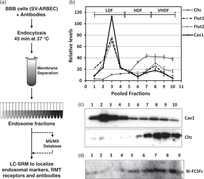
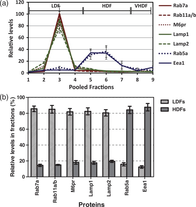
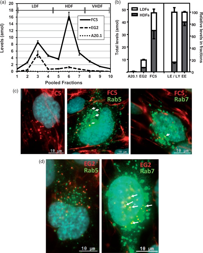
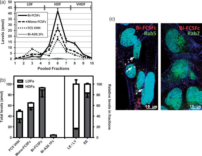
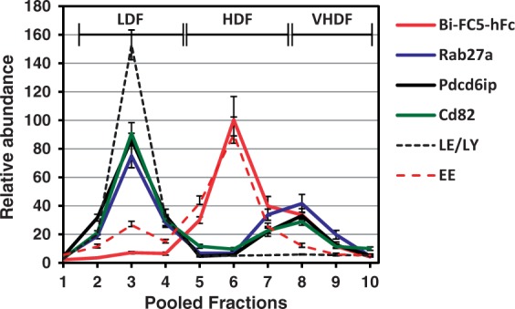
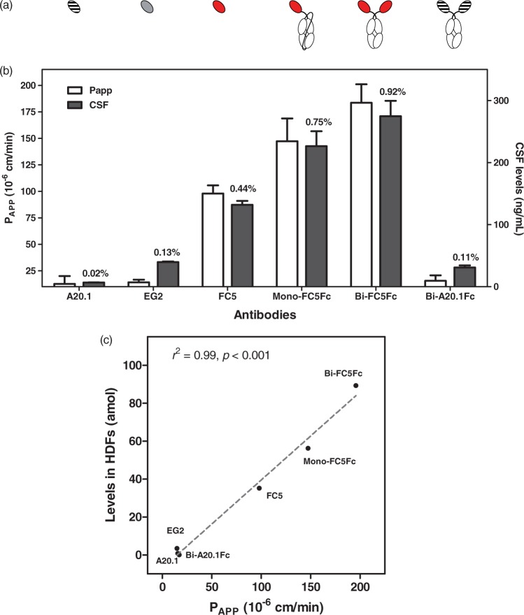
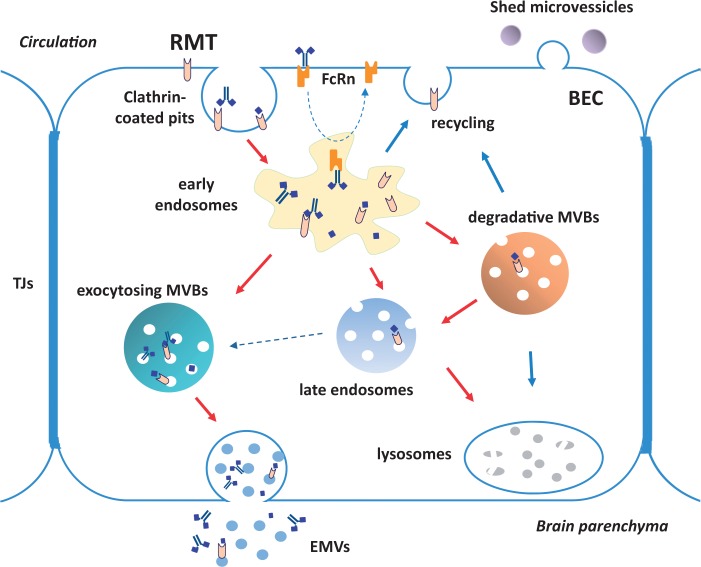
Current methods for examining antibody trafficking are either non-quantitative such as immunocytochemistry or require antibody labeling with tracers. We have developed a multiplexed quantitative method for antibody 'tracking' in endosomal compartments of brain endothelial cells. Rat brain endothelial cells were co-incubated with blood-brain barrier (BBB)-crossing FC5, monovalent FC5Fc or bivalent FC5Fc fusion antibodies and control antibodies. Endosomes were separated using sucrose-density gradient ultracentrifugation and analyzed using multiplexed mass spectrometry to simultaneously quantify endosomal markers, receptor-mediated transcytosis (RMT) receptors and the co-incubated antibodies in each fraction. The quantitation showed that markers of early endosomes were enriched in high-density fractions (HDF), whereas markers of late endosomes and lysosomes were enriched in low-density fractions (LDF). RMT receptors, including transferrin receptor, showed a profile similar to that of early endosome markers. The in vitro BBB transcytosis rates of antibodies were directly proportional to their partition into early endosome fractions of brain endothelial cells. Addition of the Fc domain resulted in facilitated antibody 'redistribution' from LDF into HDF and additionally into multivesicular bodies (MVB). Sorting of various FC5 antibody formats away from late endosomes and lysosomes and into early endosomes and a subset of MVB results in increased antibody transcytosis at the abluminal side of the BBB.

Keywords: Intracellular trafficking; blood–brain barrier; mass spectrometry; selected reaction monitoring.


Figures

Figure 1.
Figure 1.
BBB endosome isolation and SRM analysis. (a) Workflow outlining the method for isolation of endosomes for analysis with LC-SRM. Details are described in Methods section. (b) Relative levels clathrin (Cltc), caveolin-1 (Cav1) and flotilin-1 (Flot1) and -2 (Flot2), markers various types of endocytic vesicles (clathrin-coated pits, caveolae and clathrin-independent endocytic vesicles, respectively) in endosomal fractions of BBB SV-ARBEC cells exposed to 5 mg/ml of Bi-FC5Fc for 45 min. The levels are determined using multiplexed LC-SRM. Shown are relative abundance (mean ± SD) of protein-specific peptides values from three endosome preparations. Fractions 1–4 are designated low-density fractions (LDFs); fractions 4–7 high-density fractions (HDFs); fractions 7–10 vHDFs. (c) Levels of clathrin and caveolin-1 in the same endosomal fractions detected by Western blot. (d) Levels of bivalent BBB-crossing antibody Bi-FC5Fc in the same endosomal fractions detected by Western blot.
Figure 2.
Figure 2.
(a) Relative levels of markers of late endosomes (Rab7a, Rab11a/b), lysosomes (Lamp1/2, M6pr) and early endosomes (Eea1, Rab5a) among endosome fractions of BBB cells, measured using LC-SRM. Peptides identified for Rab11 are common between Rab11a and Rab11b. M6pr is cation-dependent mannose-6-phosphate receptor. Shown are relative abundance (mean ± SD) of protein-specific peptides values from at least three endosome preparations. (b) Percentage of the same makers in LDFs and HDFs relative to total fractions as analyzed by LC-SRM. Shown are mean ± SD from at least three independent endosome preparations.
Figure 3.
Figure 3.
LC-SRM quantification and fluorescent tracking of VHH levels in SV-ARBEC endosomes. Cells were exposed to a mixture of VHHs, each at 5 µg/ml, for 45 min, prior to endosome fractionation as described in Materials and Methods. (a) Absolute levels of FC5, EG2 and A20.1 in endosome fractions of BBB cells measured using LC-SRM. Absolute levels are presented in attomoles (amol) corrected to 1 µg of total endosomal protein from at least three endosome preparations (mean ± SD). Fractions corresponding to LDFs and HDFs are indicated. (b) Levels of VHHs in LDFs and HDFs as determined by LC-SRM. Total levels of the three VHHs are shown on the left and the mean percentage of the markers of late endosomes/lysosomes (LE/LY) and early endosomes (EE) are shown on the right. Shown are mean ± SD from three independent endosome preparations. LE/LY markers include M6pr, Rab 7,11 and Lamp 1, 2. EE markers include Rab 5 a, Eea1. (c) Internalization of Cy5.5-labeled FC5 into non-transduced SV-ARBEC cells (left panel) and its co-localization (white arrows) with endosome markers in RFP-Rab5 (middle panel) or RFP-Rab7 (right panel) transduced SV-ARBEC. (d) Internalization and co-localization (white arrows) of Cy5.5-labeled EG2 with endosome markers in RFP-Rab5 (left panel) and RFP-Rab7 (right panel) transduced BEC. Cells were transduced and internalization studies performed as described in Materials and Methods. Micrographs are representative of three independent experiments.
Figure 4.
Figure 4.
LC-SRM quantification and fluorescent tracking of VHH-Fc fusion proteins in SV-ARBEC endosomes. (a) Absolute levels of FC5 VHH, Mono-FC5Fc, Bi-FC5Fc and Bi-A20.1Fc in endosome fractions of SV-ARBEC cells incubated separately with each of the antibodies (5 µg/ml, 45 min) and measured using LC-SRM. Absolute levels are presented in attomoles (amol) corrected to 1 µg of total endosomal protein from at least three endosome preparations (mean ± SD). Fractions corresponding to LDFs and HDFs are also indicated. (b) Levels in LDFs and HDFs as determined by LC-SRM. Total levels of the three antibodies are shown on the left and the mean percentage of the markers of late endosomes/lysosomes (LE/LY) and early endosomes (EE) are shown on the right. Shown are mean ± SD from three independent endosome preparations. LE/LY markers include M6pr, Rab 7,11 and Lamp 1, 2. EE markers include Rab 5a, Eea1. (c) Co-localization (white; indicated by white arrows) of Cy5.5-labeled Bi-FC5Fc (red) with endosome markers in RFP-Rab5 (left panel) and RFP-Rab7 (right panel) (both in green) – transduced BEC. Actin filaments labeled with Alexa Fluor 488 Phalloidin are shown in blue. Nuclei are labeled with Hoechst (shown in turquoise). Cells were transduced and internalization studies performed as described in Materials and Methods. Micrographs are representative of three independent experiments.
Figure 5.
Figure 5.
Relative levels of Bi-FC5Fc and multivesicular body (MVB)-associated proteins Rab27a, Pdcd6ip and Cd82 among endosome fractions of SV-ARBEC cells incubated with Bi-FC5Fc (5 µg/ml, 45 min). Also shown are average profiles (mean ± SD from three independent endosome preparations) of the markers of late endosomes/lysosomes (LE/LY) and early endosomes (EE) described in Figure 1.
Figure 6.
Figure 6.
In vitro and in vivo BBB crossing of examined antibodies. (a) Structural representation of tested antibodies. (b) Apparent permeability coefficient (Papp, left axis) and CSF levels (right axis) of the tested antibodies. Papp values were derived from in vitro BBB assay as described in Materials and Methods. CSF levels were measured after iv. administration of a single 7 mg/kg dose of each antibody to rats. CSF levels of single-domain antibodies (A20.1, EG2, FC5) and Fc fusion molecules (Mono-FC5Fc, Bi-FC5Fc) were determined 30 min and 24 h after iv injection, respectively (combined data from Haqqani et al. and Farrington et al.). CSF/serum ratios are indicated above the corresponding bars. Results are mean ± SD from at least four experiments. (C) Correlation between the in vitro BBB permeability coefficient (Papp) and levels of six antibodies examined in this study in HDFs of BEC endosomes.
Figure 7.
Figure 7.
A schematic diagram of the proposed mechanisms/pathways of intracellular sorting and trafficking of the BBB-crossing antibody FC5. FC5 (monomeric or Fc fusion) internalizes into BEC via clathrin-coated vesicles and is initially sorted to early endosomes. FC5 fusion to Fc fragment (mono- or bi-valent) also triggers FcRn-mediated recycling and further re-directs the antibody away from lysosomal degradation. At least partial dissociation of FC5 from its receptor likely occurs in early endosomes under acidic pH, and the receptor is recycled back to plasma membrane. Monomeric FC5 is partially sorted to late endosomes and likely via degradative MVBs into lysosomes; however, a major portion of monomeric FC5 and virtually all of internalized bi-valent FC5Fc fusion are ‘retained’ in early endosomes and are released on the abluminal side of BEC, either as free antibody or via EMVs.

References

    1. Abbott NJ, Patabendige AAK, Dolman DEM, et al. Structure and function of the blood-brain barrier. Neurobiol Dis 2010; 37: 13–25. - PubMed
    1. Pardridge WM. Drug and gene delivery to the brain: the vascular route. Neuron 2002; 36: 555–558. - PubMed
    1. Jones AR, Shusta EV. Blood-brain barrier transport of therapeutics via receptor-mediation. Pharm Res 2007; 24: 1759–71. - PMC - PubMed
    1. Pardridge WM, Buciak JL, Friden PM. Selective transport of an anti-transferrin receptor antibody through the blood-brain barrier in vivo. J Pharmacol Exp Ther 1991; 259: 66–70. - PubMed
    1. Yu YJ, Zhang Y, Kenrick M, et al. Boosting brain uptake of a therapeutic antibody by reducing its affinity for a transcytosis target. Sci Transl Med 2011; 3: 84ra44. - PubMed