

Ginsenoside Rg3 inhibits angiogenesis in a rat model of endometriosis through the VEGFR-2-mediated PI3K/Akt/mTOR signaling pathway
- PMID: 29140979
- PMCID: PMC5687597
- DOI: 10.1371/journal.pone.0186520
Ginsenoside Rg3 inhibits angiogenesis in a rat model of endometriosis through the VEGFR-2-mediated PI3K/Akt/mTOR signaling pathway
Abstract
Objective: This study aimed to investigate the link between the inhibitory effect of ginsenoside Rg3 on the ectopic endometrium growth and the VEGFR-2-mediated PI3K/Akt/mTOR signaling pathway, a mechanism known to inhibit angiogenesis and induce ectopic endometrial cell apoptosis.
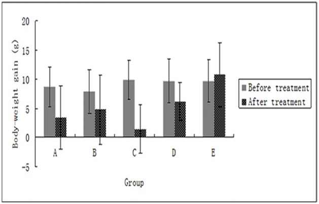
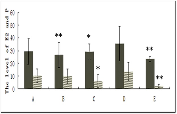
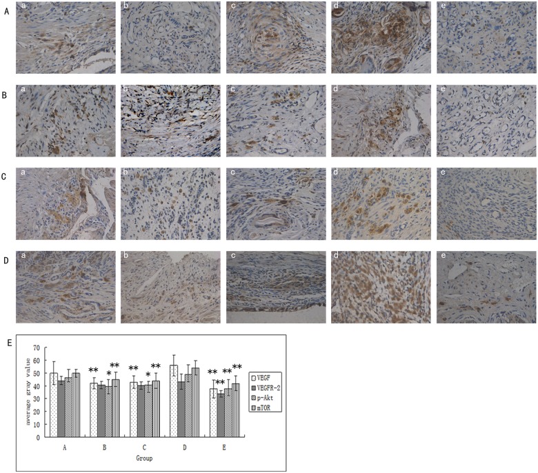
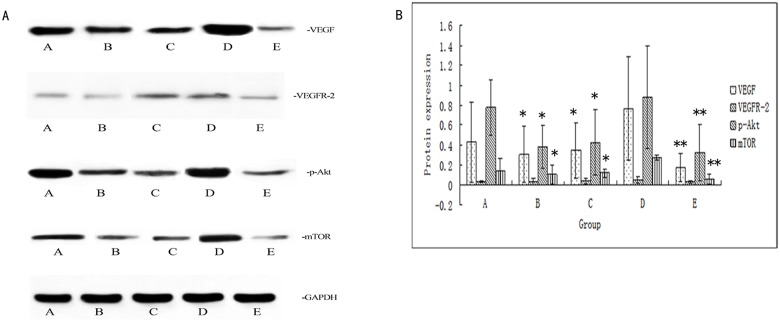
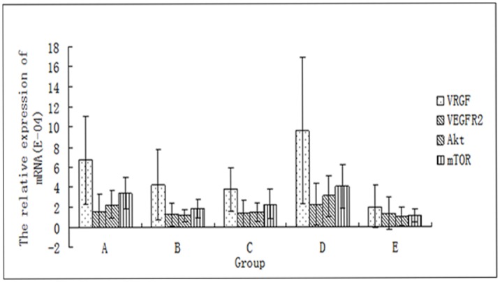
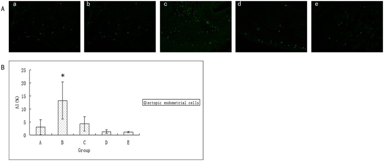
Materials and methods: A model of endometriosis was established by allotransplantation in rats. The rats were randomly divided into 5 groups: the ginsenoside Rg3 low-dose group (group A,5mg/kgBW/d of ginsenoside Rg3), the ginsenoside Rg3 high-dose group (group B, 10mg/kgBW/d of ginsenoside Rg3), the gestrinone group (group C, 0.5mg/kgBW/d of gestrinone), the control group (groupD, 10ml/kg BW/d of 0.5%CMC-Na) and the ovariectomized group (group E, 10ml/kgBW/d of 0.5%CMC-Na). Rats were executed after 21 days of continuous administration. The ectopic endometrium volume was measured and the inhibitory rate was calculated. The levels of serum estradiol (E2) and progesterone (P) were detected by Electro-Chemiluminescence Immunoassay (ECLI). The protein expressionof VEGF, VEGFR-2, p-Akt, and p-mTOR inthe ectopic endometrium wastested by immunohistochemistry(IHC) and Western Blotting. The mRNA expression levels of VEGF, VEGFR-2, Akt, and mTOR were tested by Real-Time Polymerase Chain Reaction (PCR). The apoptosis rate of the ectopic endometrial cells was detected by Terminal Deoxynucleotidyl Transferase-mediated Digoxigenin-dUTP Nick-End Labeling Assay(TUNEL).
Main results: Tissue measurements revealed a dose-dependent inhibition effect of ginsenoside Rg3 on the growth of the ectopic endometrium in treated rats compared to controls. Immunohistochemical and Western Blotting assays confirmed that the expression of VEGF, p-Akt, and p-mTOR was down-regulated in ginsenoside Rg3 -treated lesions. Real-time PCR results also showed that the mRNA expression levels of VEGF, Akt, and mTOR in the ectopic endometrium were reduced.
Conclusions: The present study demonstrates, for the first time, that ginsenoside Rg3 suppresses angiogenesis in developing endometrial lesions. The ginsenoside Rg3 inhibitory effect on the growth of the ectopic endometrium in EMs rats might occur through the blocking of the VEGFR-2-mediated PI3K/Akt/mTOR signaling pathway, thus halting angiogenesis and promoting the apoptosis of ectopic endometrial cells.
Conflict of interest statement
Figures

References
-
- Kodaman PH. Current strategies for endometriosis management.Obstet Gynecol Clin North Am.2015;42(1): 87–101. doi: 10.1016/j.ogc.2014.10.005 - DOI - PubMed
-
- Guo SW. Recurrence of endometriosis and its control. Hum Reprod Update. 2009;15(4): 441–461. doi: 10.1093/humupd/dmp007 - DOI - PubMed
-
- Matsumoto T, Yamazaki M, Takahashi H, Kajita S, Suzuki E, Tsuruta T,et al. Distinct β-catenin and PIK3CA mutation profiles in endometriosis-associated ovarian endometrioid and clear cell carcinomas. Am J Clin Pathol. 2015;144(3):452–452. doi: 10.1309/AJCPZ5T2POOFMQVN - DOI - PubMed
-
- Wang N, Liu B, Liang L, Wu Y, Xie H, Huang J,et al. Antiangiogenesis therapy of endometriosis using PAMAM as a gene vector in a noninvasive animal model. Biomed Res Int. 2014;2014:546479 doi: 10.1155/2014/546479 - DOI - PMC - PubMed
-
- Carvalho Mde S, Pereira AM, Martins JA, Lopes RC.Predictive factors for recurrence of ovarian endometrioma after laparoscopic excision. Rev Bras Ginecol Obstet. 2015;37(2): 77–81. doi: 10.1590/SO100-720320140005199 - DOI - PubMed
MeSH terms
Substances
LinkOut - more resources
Full Text Sources
Other Literature Sources
Medical
Miscellaneous
