. 2017 Nov 15;12(11):e0187014.
doi: 10.1371/journal.pone.0187014. eCollection 2017.

Wavelet brain angiography suggests arteriovenous pulse wave phase locking

Affiliations

Wavelet brain angiography suggests arteriovenous pulse wave phase locking

William E Butler. PLoS One. .

Abstract

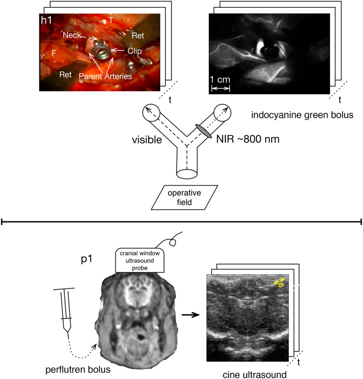
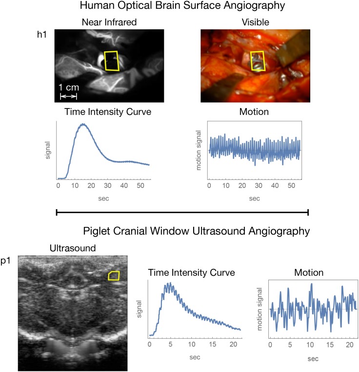
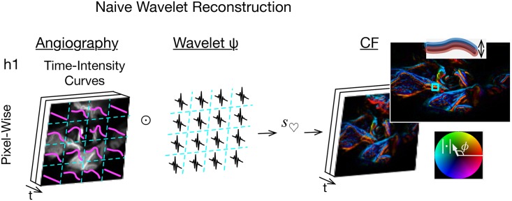
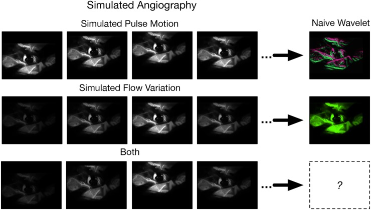
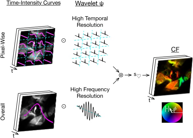
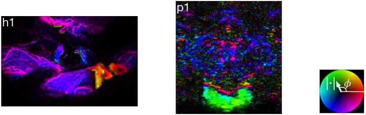
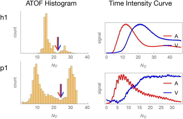
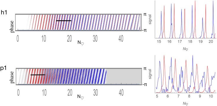
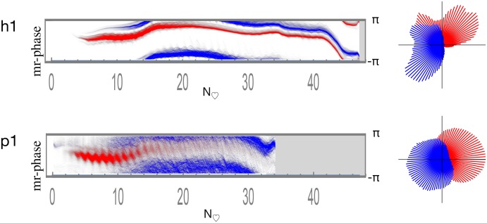
When a stroke volume of arterial blood arrives to the brain, the total blood volume in the bony cranium must remain constant as the proportions of arterial and venous blood vary, and by the end of the cardiac cycle an equivalent volume of venous blood must have been ejected. I hypothesize the brain to support this process by an extraluminally mediated exchange of information between its arterial and venous circulations. To test this I introduce wavelet angiography methods to resolve single moving vascular pulse waves (PWs) in the brain while simultaneously measuring brain pulse motion. The wavelet methods require angiographic data acquired at significantly faster rate than cardiac frequency. I obtained these data in humans from brain surface optical angiograms at craniotomy and in piglets from ultrasound angiograms via cranial window. I exploit angiographic time of flight to resolve arterial from venous circulation. Initial wavelet reconstruction proved unsatisfactory because of angiographic motion alias from brain pulse motion. Testing with numerically simulated cerebral angiograms enabled the development of a vascular PW cine imaging method based on cross-correlated wavelets of mixed high frequency and high temporal resolution respectively to attenuate frequency and motion alias. Applied to the human and piglet data, the method resolves individual arterial and venous PWs and finds them to be phase locked each with separate phase relations to brain pulse motion. This is consistent with arterial and venous PW coordination mediated by pulse motion and points to a testable hypothesis of a function of cerebrospinal fluid in the ventricles of the brain.


Conflict of interest statement

Competing Interests: WEB has applied for a patent on the method of wavelet angiography: There is a patent submission USPTO 62187631 Wavelet Reconstruction of Moving Brain Pulse on the wavelet angiography method employed to produce the data that is then subjected to further analysis in this paper. There are no declarations related to employment, consultancy, products in development, etc. This does not alter my adherence to PLOS ONE policies on sharing data and materials.

Figures

Fig 1
Fig 1. Angiography sampled faster than cardiac rate.
Upper panel is human brain surface optical angiography with simultaneous visible recording via a beam splitter. The aneurysm is of the right middle cerebral artery, where F = frontal lobe, T = temporal lobe, and Ret = retractors. The lower panel is piglet ultrasound angiography via cranial window.
Fig 2
Fig 2. Representative snapshot images and temporal data summaries for human subject h1 and piglet subject p1.
The yellow polygon in the images gives the region for motion tracking.
Fig 3
Fig 3. Naive wavelet angiography and motion alias.
Apply wavelet transforms (⊙) to the pixel-wise time signals, filter for cardiac wavelet scale (s), and render by a brightness-hue color model that represents CF magnitude as brightness and phase as hue (right bottom inset). The double inset top right shows motion alias in the lengthwise bimodal phase.
Fig 4
Fig 4. Optical angiogram simulation.
The top row is simulated pulse motion. The bottom row is simulated intraluminal contrast variation. The right column shows an image after naive CF wavelet filtering. The simulated pulse motion and intraluminal contrast variation can be combined.
Fig 5
Fig 5. Cross-correlated wavelet angiography.
Top left, apply high temporal resolution wavelet (⊙) transformation to the pixel-wise time intensity curves. Bottom left, apply high frequency resolution wavelet transformation to the overall time intensity curve. Right side, cross-correlate these pixel-wise (⊗), filter for cardiac wavelet scale (s), inverse wavelet transform, then cine render.
Fig 6
Fig 6. Wavelet angiography snapshots.
Fig 7
Fig 7. Arteriovenous classification.
Arterial and venous pixels are separated according to angiographic time of flight (ATOF). The derived time intensity curves are respectively consistent with predominantly arterial and venous temporal behavior.
Fig 8
Fig 8. Arterial and venous time-indexed phase histograms with line scans.
Pixels classified by ATOF as arterial are in red and those as venous in blue. Relative histogram count is represented as brightness. The horizontal axis is time is depicted in heartbeats to facilitate human-piglet comparison. The vertical axis is phase ranging as [−π, π]. There is a consistent arteriovenous phase difference across the bolus passage.
Fig 9
Fig 9. Arterial and venous motion-referenced phase histograms.
Pixels classified by ATOF as arterial are in red and those as venous in blue. Relative histogram count is represented as brightness. The horizontal axis is time is depicted in heartbeats to facilitate human-piglet comparison. The vertical axis is phase ranging as [−π, π]. Arterial versus venous phase appears approximately fixed in difference across the entire bolus passage. The circular phase histograms on the right are calculated by summing the time-indexed phase histograms on the left across time.

Similar articles

Cited by

References

    1. Wilson MH. Monro-Kellie 2.0: The dynamic vascular and venous pathophysiological components of intracranial pressure. Journal of Cerebral Blood Flow & Metabolism. 2016;36(8):1338–1350. doi: 10.1177/0271678X16648711 - DOI - PMC - PubMed
    1. Tomita M, Osada T, Schiszler I, Tomita Y, Unekawa M, Toriumi H, et al. Automated method for tracking vast numbers of FITC-labeled RBCs in microvessels of rat brain in vivo using a high-speed confocal microscope system. Microcirculation (New York, NY: 1994). 2008;15(2):163–174. doi: 10.1080/10739680701567089 - DOI - PubMed
    1. Butler WE, Agarwalla PK, Codd P. CSF in the Ventricles of the Brain Behaves as a Relay Medium for Arteriovenous Pulse Wave Coupling. PloS One. - PMC - PubMed
    1. Nyqvist H. Certain topics in telegraph transmission theory. Transactions of the AIEE. 1928;47:617–644.
    1. Shannon CE. Communication in the Presence of Noise. Proceedings of the Institute of Radio Engineers. 1949;37(1):10–21.