. 2018 Dec;46(8):1659-1670.
doi: 10.1080/21691401.2017.1388249. Epub 2017 Nov 16.

Myocardial reparative functions of exosomes from mesenchymal stem cells are enhanced by hypoxia treatment of the cells via transferring microRNA-210 in an nSMase2-dependent way

Affiliations

Myocardial reparative functions of exosomes from mesenchymal stem cells are enhanced by hypoxia treatment of the cells via transferring microRNA-210 in an nSMase2-dependent way

Jinyun Zhu et al. Artif Cells Nanomed Biotechnol. 2018 Dec.

Abstract

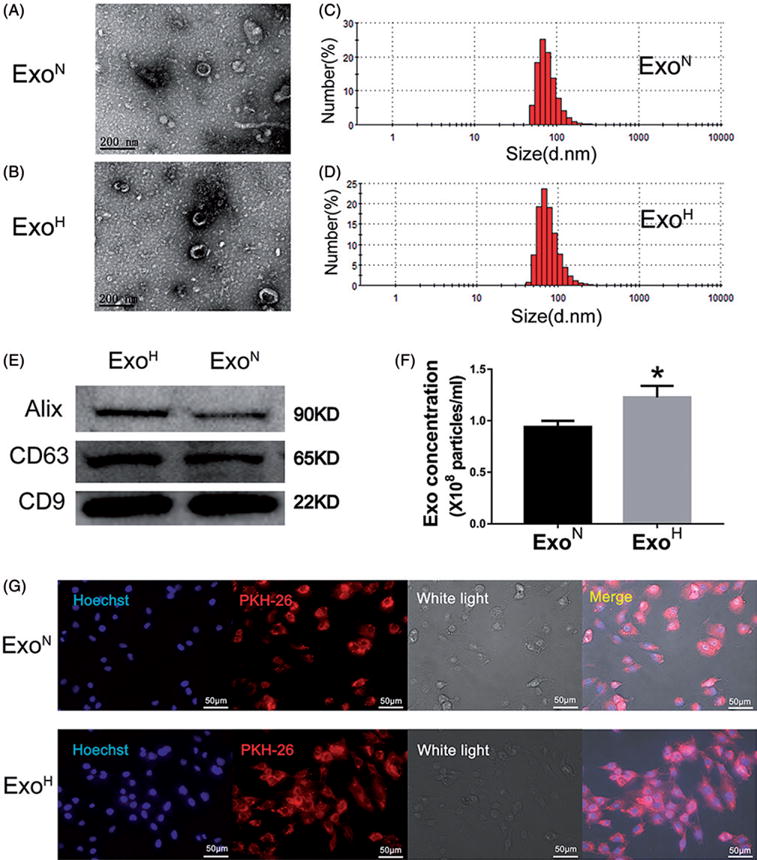
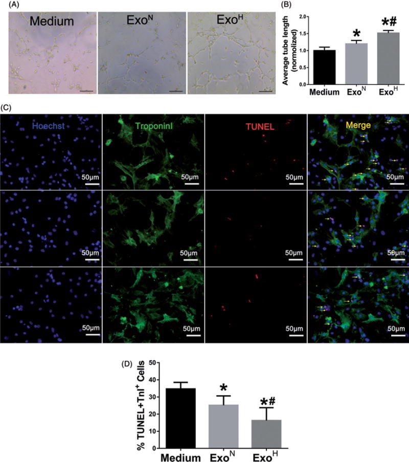
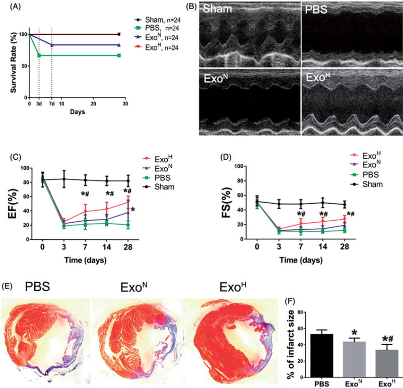
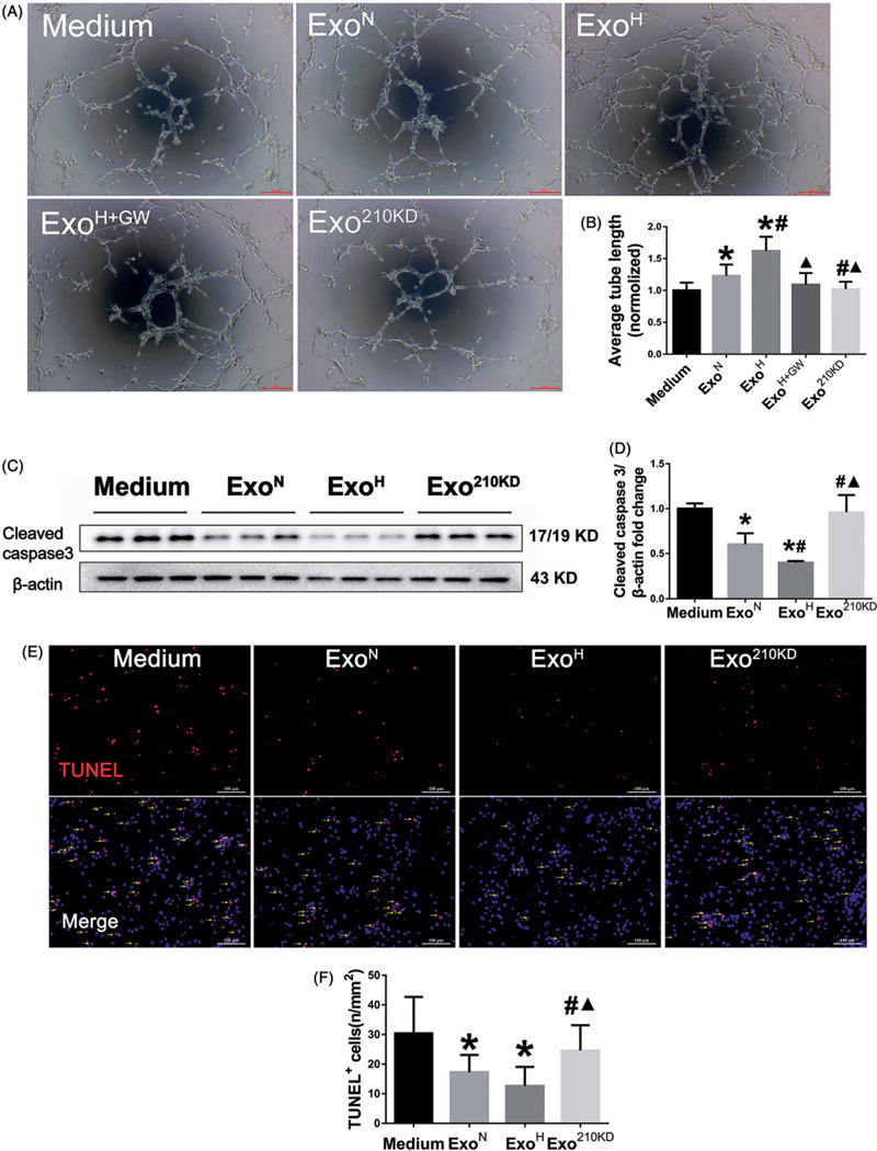
Hypoxia treatment enhances paracrine effect of mesenchymal stem cells (MSCs). The aim of this study was to investigate whether exosomes from hypoxia-treated MSCs (ExoH) are superior to those from normoxia-treated MSCs (ExoN) for myocardial repair. Mouse bone marrow-derived MSCs were cultured under hypoxia or normoxia for 24 h, and exosomes from conditioned media were intramyocardially injected into infarcted heart of C57BL/6 mouse. ExoH resulted in significantly higher survival, smaller scar size and better cardiac functions recovery. ExoH conferred increased vascular density, lower cardiomyocytes (CMs) apoptosis, reduced fibrosis and increased recruitment of cardiac progenitor cells in the infarcted heart relative to ExoN. MicroRNA analysis revealed significantly higher levels of microRNA-210 (miR-210) in ExoH compared with ExoN. Transfection of a miR-210 mimic into endothelial cells (ECs) and CMs conferred similar biological effects as ExoH. Hypoxia treatment of MSCs increased the expression of neutral sphingomyelinase 2 (nSMase2) which is crucial for exosome secretion. Blocking the activity of nSMase2 resulted in reduced miR-210 secretion and abrogated the beneficial effects of ExoH. In conclusion, hypoxic culture augments miR-210 and nSMase2 activities in MSCs and their secreted exosomes, and this is responsible at least in part for the enhanced cardioprotective actions of exosomes derived from hypoxia-treated cells.

Keywords: Exosomes; MSCs; hypoxia; microRNA210; myocardial infarction; nSMase2.


Conflict of interest statement

Disclosure statement

No potential conflict of interest was reported by the authors.

Figures

Figure 1
Figure 1
Characterization and functional validation of exosomes derived from mesenchymal stem cells (MSCs). (A,B) Cup-shaped morphology of purified exosomes assessed by TEM. Scale bar =200 nm. (C,D) DLS analysis of ExoN and ExoH shows that both exosomes had the typical size range (30–120 nm). (E) Exosomal markers assessed by Western blotting. (F) The number of exosomal particles assessed by NTA. *p < .05, vs. ExoN. (G) Uptake of red fluorescence dye PKH26 labelled exosomes into H9C2s. Scale bar =50 μm.
Figure 2
Figure 2
ExoH enhances angiogenic activity and survive of recipient cells. (A) Representative images showing tube formation in HUVECs treated with Medium, ExoN or ExoH. Bar =100 μm. (B) Quantification of tube length in each group (n = 12). (C) TUNEL staining for apoptosis of CMs after treated with Medium (top), ExoN (middle), and ExoH (bottom) and exposure to hypoxia/reoxygenation. Arrows point cells that are positive for both TUNEL (red) and Troponin I (green). Blue: Hoechst. Bar =50 μm. (D) Apoptotic cells were quantified as the percentage of CMs that were positive for TUNEL staining (n = 12), *p < .05, vs. Medium; #p < .05, vs. ExoN.
Figure 3
Figure 3
Augmenting cardiac function and ameliorating fibrosis after MI by ExoH. (A) The number of survival animals was recorded for each day of the study period (from 0 to 28 d). (B) Representative echocardiography images showing significantly increased wall motion in ExoH treated hearts. (C,D) Gradually increased ejection fraction (EF) and fractional shortening (FS) in mice transplanted with ExoH compared with other groups (n = 24 for Sham, 16 for PBS, 20 for ExoN and 24 for ExoH). (E) The cross-sections of infarcted hearts were analysed with Masson trichrome staining at 4 weeks after infarction. The fibrosis in the scar of infarcted hearts was shown in blue. (F) The fibrotic scar areas were quantified (n = 6). *p < .05, vs. PBS; #p < .05, vs. ExoN.
Figure 4
Figure 4
ExoH promotes angiogenesis and reduces cardiac apoptosis after infarction. (A) Capillary at the border zone on day 28 after MI was identified by staining with CD31 (red) and nuclei (blue). (B) Quantification of CD31+ cells in A (n = 8). (C) Arterioles at the border zone on day 28 after MI were identified by staining with α-SMA (green) and nuclei (blue). (D) Quantification of α-SMA+ cells in C (n = 8). (E) Quantification of TUNEL+ cells in Supplemental Figure S3(A). Apoptosis rate was quantified as the percentage of cells that were positive for TUNEL staining. (F) Quantification of Sca-1+ cells in Supplemental Figure S3(B) (n = 6). *p < .05, vs. PBS; #p < .05, vs. ExoN. Scale bar =100 μm.
Figure 5
Figure 5
miR-210 mediates the improved biological function of recipient cells. (A) Tube formation assay using HUVECs treated with Medium, ExoN, ExoH, ExoH+GW or Exo210 KD. Scale bar =100 μm. (B) Quantification of tube length in A (n = 6). (C) Western blot for cleaved caspase3 protein in MSCs treated with different exosomes. (D) Quantification of cleaved caspase3 in C (n = 6). (E) TUNEL staining for apoptosis of CMs after treated with exosomes and exposure to hypoxia/reoxygenation. Arrows point cells that are positive for TUNEL. Scale bar = 100μm. (F) Quantification of apoptotic cells in E (n = 6). The number of apoptotic cells was increased in ExomiR210 KD group. *p <.05, vs. Medium; #p<.05, vs. ExoN; p<.05, vs. ExoH.
Figure 6
Figure 6
nSMase2 modulates miR-210 salutary effects under hypoxia condition. (A) RT-PCR analysis of Rab27a and SMPD3 mRNA in MSCs after normoxia (Control) or hypoxia (HP) treatment (n = 6). (B) Western blot analysis of nSMase2 protein in MSCs (*p < .05, vs. Control). (C,D) Quantification of miR-210 by RT-PCR in conditioned medium (C) and exosomes (D) from MSCs treated with different dose of GW4869 under normoxia (Control) and hypoxia (HP) conditions (n = 6, p < .05, vs. 0 μM; #p < .05, vs. 5 μM). (E) Western blot analysis of apoptotic maker in H9C2s treated with Medium, ExoN, ExoH or ExoH+GW. (F) Quantification of cleaved caspase3
Figure 7
Figure 7
HIF-1α modulated nSMase2 expression in MSCs. (A) Western blot assay shows that stabilization of HIF-1α by DMOG increased nSMsase2 expression in MSCs under normoxia condition. (B) Quantification of HIF-1α and nSMase2 proteins in A (n = 3, repeated three times. *p < .05, vs. Control). (C) Western blot analysis of HIF-1α and nSMsase2 in MSCs that were incubated with two siRNA directed against HIF-1α (HIF-1α siRNA) or scrambled control as negative control (NC) and then subjected to hypoxia for 6 hours. (D) Quantification of HIF-1α and nSMase2 proteins in C. (n = 4, repeated two times. *p < .05, vs. NC).
Figure 8
Figure 8
Schematic representation of the effects and mechanisms of hypoxic-MSCs derived exosomes for cardiac repair after myocardial infarction. Hypoxia induced stabilization and nucleus translocation of HIF-1α causing the activation of downstream target nSMase2 (gene SMPD3), presumably through binding putative HREs in the promoter sequence of SMPD3 gene (The dotted line shows the hypothesis which is not confirmed). Multivesicular Bodies (MVBs) are late endosomal compartments that contain intraluminal vesicles as exosomes formed from the inward invagination of endosomal membranes, which requires ceramide generation by nSMase2. Increased miR-210 by hypoxia is encapsulated in the exosomes in an nSMase2 dependent way. Hypoxic-MSCs secreted exosomes deliver miR-210 to the infarct heart, resulting in modulation of angiogenesis, survival and migration of cardiac cells that ultimately leads significant augmentation of cardiac regeneration in the heart after MI.

References

    1. Lee CY, Kim R, Ham O, et al. Therapeutic potential of stem cells strategy for cardiovascular diseases. Stem Cell Int. 2016;2016:4285938. - PMC - PubMed
    1. Ahuja P, Perriard E, Perriard JC, et al. Sequential myofibrillar breakdown accompanies mitotic division of mammalian cardiomyocytes. J Cell Sci. 2004;117:3295–3306. - PubMed
    1. Singla DK. Stem cells and exosomes in cardiac repair. Curr Opin Pharmacol. 2016;27:19–23. - PubMed
    1. Kochegarov A, Lemanski LF. New trends in heart regeneration: a review. J Stem Cells Regen Med. 2016;12:61–68. - PMC - PubMed
    1. Marote A, Teixeira FG, Mendes-Pinheiro B, et al. MSCs-derived exosomes: cell-secreted nanovesicles with regenerative potential. Front Pharmacol. 2016;7:231. - PMC - PubMed