Review
. 2017 Nov 15;43(1):101.
doi: 10.1186/s13052-017-0419-z.

Congenital myopathies: clinical phenotypes and new diagnostic tools

Collaborators, Affiliations
Review

Congenital myopathies: clinical phenotypes and new diagnostic tools

Denise Cassandrini et al. Ital J Pediatr. .

Abstract

Congenital myopathies are a group of genetic muscle disorders characterized clinically by hypotonia and weakness, usually from birth, and a static or slowly progressive clinical course. Historically, congenital myopathies have been classified on the basis of major morphological features seen on muscle biopsy. However, different genes have now been identified as associated with the various phenotypic and histological expressions of these disorders, and in recent years, because of their unexpectedly wide genetic and clinical heterogeneity, next-generation sequencing has increasingly been used for their diagnosis. We reviewed clinical and genetic forms of congenital myopathy and defined possible strategies to improve cost-effectiveness in histological and imaging diagnosis.

Keywords: Congenital myopathy; Muscle MRI; Muscle biopsy; Next generation sequencing.


Conflict of interest statement

Authors' information

#The Italian Network on Congenital Myopathies is made of the following researchers:

Angela Berardinelli1, Enrico S. bertini 2, Giacomo Comi3, Adele D’Amico2, Maria Alice Donati4, Maria Teresa Dotti5, Fabiana Fattori2, Marina Grandis6, Lorenzo Maggi7, Francesca Magri3, Maria A. Maioli8, Alessandro Malandrini5, Francesco Mari4, Roberto Massa9, Eugenio Mercuri10, Luciano Merlini11, Maurizio Moggio12, Marina Mora7, Lucia O. Morandi7, Olimpia Musumeci13, Vincenzo Nigro14, Marika Pane10, Elena Pegoraro15, Elena M. Pennisi16, Lorenzo Peverelli12, Giulia Ricci17, Carmelo Rodolico13, Lucia Ruggiero18, Michele Sacchini4, Lucio Santoro18, Marco Savarese14, Gabriele Siciliano17, Alessandro Simonati19, Paola Tonin19, Antonio Toscano13.

1 UO Neuropsichiatria Infantile, Ist. Mondino, Pavia, 2 Lab. of Molecular Medicine for Neuromuscular and Neurodegenerative Disorders Bambino Gesù Children’s Hospital, Rome, 3 UO Neurology, Maggiore Hospital, Milan, 4 Meyer Children Hospital, Florence, 5 Neurology, University of Siena, Siena,6 1 Department of Neuroscience, Rehabilitation, Ophthalmology, Genetics, University of Genoa, Genoa, 7 Muscle Cell Biology Lab, Neuromuscular Diseases and Neuroimmunology Unit, Fondazione IRCCS C. Besta, 8 Neurophysiopathology Multiple Sclerosis Center Hospital Binaghi, Cagliari, 9 UOC Neurology, Policlinico Tor Vergata, Rome, 10 U.O. Neuropsichiatria Infantile, A. Gemelli Hospital, Rome, 11 University of Bologna, Bologna, 12 Neuromuscular and Rare Diseases Unit, Dino Ferrari Center, Milano, 13 UOC Neurology and Neuromuscular Diseases, AOU Policlinico G. Martino, Messina, 14 Telethon Institute of Genetics and Medicine, Naples, 15 Department of Neurology, Hospital of Padova, Padova, 16 UOC Neurology, San Filippo Neri, Rome, 17 UO Neurology, AOUP, Pisa, 18 UO Neurology, Policlinico Federico II, Naples, 19 UOC Neurology, Borgo Roma Hospital, Verona.

Ethics approval and consent to participate

This study was approved by the ethics committee of IRCCS Stella Maris, Pisa, Italy and other participating institutions.

Consent for publication

All the procedures complied with the Helsinki Declaration of 1975. DNA, morphological, MRI and clinical studies were performed with parental written informed consent.

Competing interests

The authors declare that they have no competing interests.

Publisher’s Note

Springer Nature remains neutral with regard to jurisdictional claims in published maps and institutional affiliations.

Figures
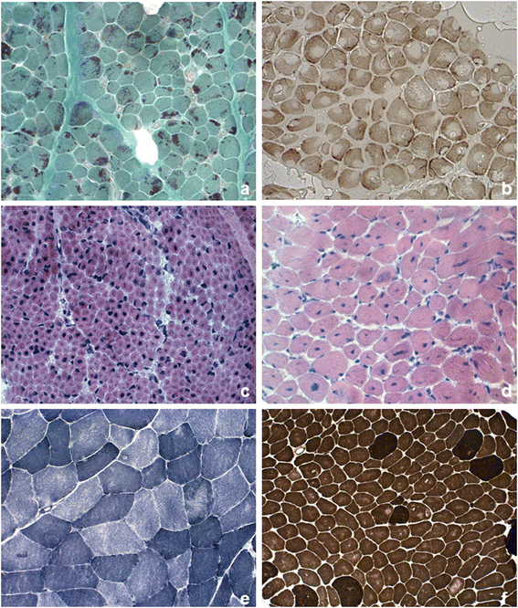
Fig. 1
Fig. 1
Pathological features in skeletal muscle biopsy observed in congenital myopathies. a Nemaline rods. Gomori trichrome stain showing clusters of purple staining rods at the periphery of most fibers and some internal within fibers. b Cores. Cytochrome c oxidase stain demonstrating numerous cores of varying size, centrally or peripherally. c Central nuclei. Quadriceps biopsy from a boy presenting X-linked myotubular myopathy. Hematoxylin–eosin stain (HE) showed large central nuclei in several fibers. d Central nuclei. Quadriceps biopsy from a 5-year-old patient with centronuclear myopathy due to a mutation in DNM2. HE demonstrated centrally placed nuclei in the majority of fibers, variation in fiber size and increased connective tissue. e Multi-minicores. NADH-TR stain showing areas in both fiber types of varying size and number devoid of oxidative enzyme stain. f Congenital fiber type disproportion. ATPase pre-incubated at pH 9,6 stain showing the small size of the light-staining type 1 fibers and type 1 fiber predominance

References

    1. North KN, Wang CH, Clarke N, Jungbluth H, Vainzof M, Dowling JJ, et al. Approach to the diagnosis of congenital myopathies. Neuromuscul Disord. 2014;24:97–116. doi: 10.1016/j.nmd.2013.11.003. - DOI - PMC - PubMed
    1. Tubridy N, Fontaine B, Eymard B. Congenital myopathies and congenital muscular dystrophies. Curr Opin Neurol. 2001;14:575–582. doi: 10.1097/00019052-200110000-00005. - DOI - PubMed
    1. Jungbluth H, Voermans NC. Congenital myopathies: not only a paediatric topic. Curr Opin Neurol. 2016;29:642–650. doi: 10.1097/WCO.0000000000000372. - DOI - PubMed
    1. Colombo I, Scoto M, Manzur AY, Robb SA, Maggi L, Gowda V, et al. Congenital myopathies: natural history of a large pediatric cohort. Neurology. 2015;84:28–35. doi: 10.1212/WNL.0000000000001110. - DOI - PMC - PubMed
    1. Fardeau M, Tome F. Congenital myopathies. In: Engler AG, Franzini-Armstrong C, editors. Myology. New York: McGraw-Hill; 1994. pp. 1487–1533.

MeSH terms