. 2017 Nov 15;19(1):121.
doi: 10.1186/s13058-017-0910-x.

Local estrogen axis in the human bone microenvironment regulates estrogen receptor-positive breast cancer cells

Affiliations

Local estrogen axis in the human bone microenvironment regulates estrogen receptor-positive breast cancer cells

Derek F Amanatullah et al. Breast Cancer Res. .

Abstract

Background: Approximately 70% of all breast cancers express the estrogen receptor, and are regulated by estrogen. While the ovaries are the primary source of estrogen in premenopausal women, most breast cancer is diagnosed following menopause, when systemic levels of this hormone decline. Estrogen production from androgen precursors is catalyzed by the aromatase enzyme. Although aromatase expression and local estrogen production in breast adipose tissue have been implicated in the development of primary breast cancer, the source of estrogen involved in the regulation of estrogen receptor-positive (ER+) metastatic breast cancer progression is less clear.

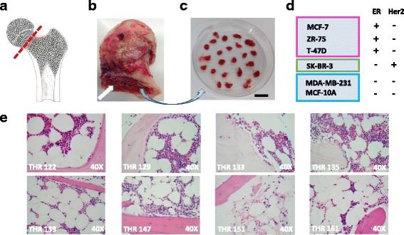
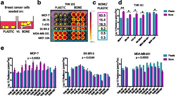
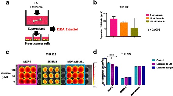
Methods: Bone is the most common distant site of breast cancer metastasis, particularly for ER+ breast cancers. We employed a co-culture model using trabecular bone tissues obtained from total hip replacement (THR) surgery specimens to study ER+ and estrogen receptor-negative (ER-) breast cancer cells within the human bone microenvironment. Luciferase-expressing ER+ (MCF-7, T-47D, ZR-75) and ER- (SK-BR-3, MDA-MB-231, MCF-10A) breast cancer cells were cultured directly on bone tissue fragments or in bone tissue-conditioned media, and monitored over time with bioluminescence imaging (BLI). Bone tissue-conditioned media were generated in the presence vs. absence of aromatase inhibitors, and testosterone. Bone tissue fragments were analyzed for aromatase expression by immunohistochemistry.

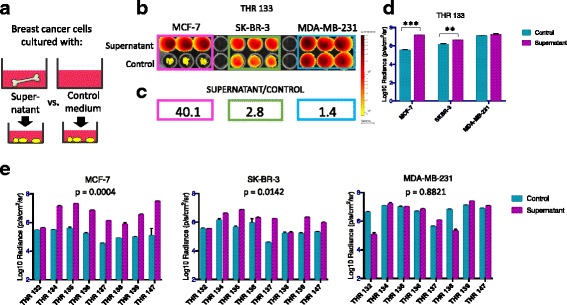
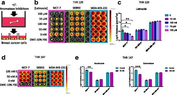
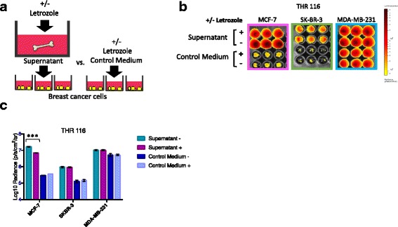
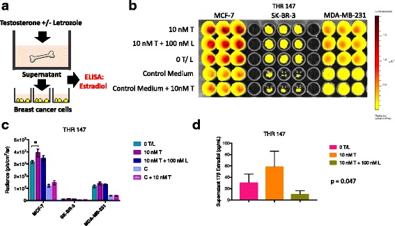
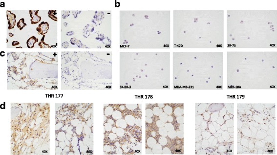
Results: ER+ breast cancer cells were preferentially sustained in co-cultures with bone tissues and bone tissue-conditioned media relative to ER- cells. Bone fragments analyzed by immunohistochemistry revealed expression of the aromatase enzyme. Bone tissue-conditioned media generated in the presence of testosterone had increased estrogen levels and heightened capacity to stimulate ER+ breast cancer cell proliferation. Pretreatment of cultured bone tissues with aromatase inhibitors, which inhibited estrogen production, reduced the capacity of conditioned media to stimulate ER+ cell proliferation.

Conclusions: These results suggest that a local estrogen signaling axis regulates ER+ breast cancer cell viability and proliferation within the bone metastatic niche, and that aromatase inhibitors modulate this axis. Although endocrine therapies are highly effective in the treatment of ER+ breast cancer, resistance to these treatments reduces their efficacy. Characterization of estrogen signaling networks within the bone microenvironment will identify new strategies for combating metastatic progression and endocrine resistance.

Keywords: Aromatase; Aromatase inhibitors; Bone tissue culture; Breast cancer metastasis to bone; Estrogen receptor positive breast cancer.


Conflict of interest statement

Ethics approval and consent to participate

Ethics approval was obtained by the Stanford University Internal Review Board (IRB). All discarded surgical specimens and patient medical record information, including diagnoses and disease history (obtained from the STRIDE database by Research IT, Stanford Medicine), were collected under IRB Protocol # 38625, with a Waiver of Consent, in accordance with regulations of the Stanford University Research Compliance Office.

Consent for publication

Not applicable.

Competing interests

The authors declare that they have no competing interests.

Publisher’s Note

Springer Nature remains neutral with regard to jurisdictional claims in published maps and institutional affiliations.

Figures

Fig. 1
Fig. 1
Human bone tissue co-culture model. a Femoral head removal during total hip replacement (THR) surgery (image credit: Moran & Rowley – Visualhistology.com). b Discarded femoral head exposing trabecular bone (white arrow). c Isolated trabecular bone tissue fragments (bar = 3 mm). d Panel of breast cancer cell lines transfected with luciferase and enhanced green fluorescence protein (EGFP): estrogen receptor-positive (ER+) MCF-7, ZR-75 and T-47D, ER-/human epidermal growth factor receptor 2 (Her2) + SK-BR-3, and ER- MDA-MB-231 and MCF-10A cells. e Hematoxylin and eosin stained histologic sections of trabecular bone tissues from a subset of THR specimens illustrating mineralized and marrow compartments
Fig. 2
Fig. 2
Human bone tissue fragments preferentially sustain estrogen receptor-positive (ER+) breast cancer cell viability in short-term co-culture. a Experimental design in which breast cancer cell suspensions were seeded into plastic wells or directly onto bone tissue fragments. b Bioluminescence imaging (BLI) of plate containing six breast cancer cell lines (ER+ MCF-7, ZR-75 and T-47D, ER-/human epidermal growth factor receptor 2 (Her2) + SK-BR-3, and ER- MDA-MB-231 and MCF-10A) on day 4 of culture shows signal generated during culture in plastic wells vs. bone tissue fragments isolated from total hip replacement (THR) specimen THR 151. c Ratio of averaged triplicate BLI signal following culture on bone vs. plastic for each cell line shown in (b), revealing highest ratios for the ER+ breast cancer cell lines MCF-7 (83.5x), ZR-75 (15.4x), and T-47D (18.3x), an intermediate ratio for ER-/HER2+ SK-BR-3 cells (9.2x), and lowest ratios for the ER- MDA-MB-231 (0.5x) and MCF-10A (0.3x) breast cells. d Averaged triplicate BLI signal detected for each cell line growing on plastic vs. bone shown in (b). BLI signal was significantly greater following culture on bone vs. plastic for the ER+ breast cancer cell lines MCF-7 (**p < 0.001), ZR-75 (*p < 0.01), T47-D (*p < 0.05), and the ER-/Her2-positive cell line SK-BR-3 (*p < 0.01), not different for the ER- MDA-MB-231 cells, and was significantly reduced for the ER- MCF-10A cells (*p < 0.05), as determined by t test (n = 3, error bars represent standard deviation). e Averaged triplicate BLI signal generated by MCF-7, SK-BR-3, and MDA-MB-231 cells cultured on plastic vs. bone fragments from a series of 12 THR surgical specimens. Signal was significantly greater following culture on bone vs. plastic for ER+ MCF-7 cells (p = 0.0053), whereas it was significantly reduced for ER- SK-BR-3 cells (p = 0.0184) and MDA-MB-231 cells (p = 0.0003) as determined by ANOVA (n = 3, error bars represent standard deviation). These results confirm that bone tissues from multiple patients preferentially sustain ER+ breast cancer cell viability in culture relative to ER- breast cancer cells
Fig. 3
Fig. 3
Conditioned media from cultured human bone tissue fragments preferentially stimulate estrogen receptor-positive (ER+) breast cancer cell proliferation. a Experimental design in which breast cancer cells were seeded into tissue culture wells containing bone tissue-conditioned supernatants vs. control medium (DMEM-10%FBS). b Bioluminescence imaging (BLI) on day 4 of culture showing signal for ER+ MCF-7, and ER- SK-BR-3 and MDA-MB-231 breast cancer cells cultured in supernatants generated with total hip replacement (THR) specimen 133 vs. control medium. c Ratio of averaged triplicate BLI signal on day 4 of culture in supernatant vs. control medium for each cell line shown in (b), revealing the highest ratio for ER+ MCF-7 (40.1x), and lower ratios for ER- SKBR-3 (2.8x) and MDA-MB-231 (1.4x) cells. d Averaged triplicate BLI signal detected for each cell line following culture in supernatant vs. control medium shown in (b). BLI signal was significantly greater during culture with bone tissue supernatants vs. control medium for ER-positive MCF-7 (***p = 0.0000028) and ER-/human epidermal growth factor receptor 2 (Her2)-positive SK-BR-3 (**p = 0.0046), but not ER- MDA-MB-231 cells, as determined by t test (n = 3, error bars represent standard deviation). e Averaged triplicate BLI signal generated by MCF-7, SK-BR-3, and MDA-MB-231 cells cultured in control vs. conditioned media generated from a series of eight THR surgical specimens. Analysis of variance demonstrated that BLI signal for cells cultured in conditioned vs. control media was dramatically increased for MCF-7 cells (p = 0.0004), moderately increased for SK-BR-3 cells (p = 0.0142), but was not changed for MDA-MB-231 cells (p = 0.8821). These results confirm that conditioned media generated from multiple patient specimens preferentially promote the proliferation of ER+ breast cancer cells
Fig. 4
Fig. 4
Bone tissue culture supernatants generated in the presence of aromatase inhibitors (AIs) have diminished capacity to promote estrogen receptor-positive (ER+) breast cancer cell proliferation. a Experimental design in which bone tissue fragments were cultured in the presence vs. absence of AIs for 48 h. b Bioluminescence imaging (BLI) signal displayed by breast cancer cells growing in the presence of conditioned media generated by bone fragments isolated from total hip replacement (THR) specimen 129 and cultured in DMEM-10%FBS plus 100 μM, 10 μM, 100 nM, and 10 nM, vs. 0 μM letrozole. c Averaged triplicate BLI signal detected for each cell line shown in (b). BLI signal was reduced for ER+ MCF-7, but not ER- SK-BR-3 or MDA-MB-231 cells cultured with bone tissue-conditioned media generated in the presence of 100 μM (**p = 0.003), 10 μM (*p = 0.016), 100 nM (*p = 0.035), and 10 nM (p = 0.056) vs. 0 μM letrozole, as determined by t test (n = 3, error bars represent standard deviation). d BLI signal displayed by breast cancer cells growing in the presence of conditioned media generated by bone fragments isolated from THR specimen 147 in the presence of 100 nM exemestane and 50 nM anastrozole vs. no agent. e Averaged triplicate BLI signal detected for cells shown in (d). BLI signal was specifically reduced for ER+ MCF-7 cells, but not ER- SK-BR-3 or MDA-MB-231 breast cancer cells cultured with bone tissue-conditioned media generated in the presence of 100 nM exemestane (***p = 0.00003) and 50 nM anastrozole (**p = 0.002) vs. no agent, as determined by t test (n = 3, error bars represent standard deviation)
Fig. 5
Fig. 5
Bone tissue culture supernatants generated in the presence of aromatase inhibitors have reduced estrogen levels and diminished capacity to promote estrogen receptor-positive (ER+) breast cancer cell proliferation. a Experimental design in which bone tissue fragments were cultured in the presence vs. absence of letrozole for 48 h. Supernatants were used to culture breast cancer cells growing on plastic, and were also analyzed for estradiol levels by ELISA. b ELISA analysis of estradiol levels in conditioned media generated by bone tissue fragments isolated from total hip replacement (THR) specimen THR 122 in the presence of 100 μM and 10 μM vs. 0 μM letrozole. The lowest estradiol levels were observed in supernatants generated in 100 μM, with higher values observed in supernatants generated in 10 μM and 0 μM letrozole, corresponding to estradiol levels of 47.5, 90.8, and 114.9 pg/mL, respectively (p < 0.0001) as determined by analysis of variance with the Brown-Forsythe test. c Bioluminescence imaging (BLI) signal detected for ER+ MCF-7, and ER- SK-BR-3 and MDA-MB-231 cells cultured in replicate supernatants generated by THR 122 fragments in the presence of 100 μM, 10 μM, and 0 μM letrozole. d Averaged triplicate BLI signal detected for each cell line shown in c. BLI signal was significantly reduced in ER+ MCF-7 cells, but not ER- SK-BR-3 or MDA-MB-231 cells cultured with conditioned media generated in the presence of 100 μM (***p = 0.0008) or 10 μM (*p = 0.03) vs. 0 μM letrozole, as determined by t test (n = 3, error bars represent standard deviation)
Fig. 6
Fig. 6
Letrozole inhibits estrogen receptor-positive (ER+) breast cancer cell proliferation indirectly by acting on bone tissues. a Experimental design for comparing relative breast cancer cell numbers during culture with bone tissue supernatants generated in the presence of letrozole vs. control medium containing letrozole. b Bioluminescence imaging (BLI) signal displayed by ER+ MCF-7, and ER- SK-BR-3 and MDA-MB-231 breast cancer cells growing in the presence of conditioned media generated by total hip replacement (THR) specimen THR 116 tissue fragments cultured in DMEM-10%FBS +/− 100 μM letrozole vs. control media (DMEM-10%FBS) +/− 100 μM letrozole. c Averaged triplicate BLI signal detected on plate shown in (b). BLI signal was specifically reduced in MCF-7, but not SK-BR-3 or MDA-MB-231 cells cultured with bone tissue culture supernatants generated in the presence of 100 μM vs. 0 μM letrozole μM (***p = 0.00003), as determined by t test (n = 3, error bars represent standard deviation). No difference was observed for any of the cell lines when 100 μM letrozole was added directly to the control medium
Fig. 7
Fig. 7
Bone tissue culture supernatants generated in the presence of testosterone have elevated estrogen levels and increased capacity to promote estrogen receptor-positive (ER+) breast cancer cell proliferation. a Experimental design in which bone tissue fragments are cultured in the presence of phenol red-free medium with 10% charcoal-stripped FBS containing testosterone in the presence vs. absence of letrozole. Supernatants were harvested and used to culture breast cancer cells growing on plastic and analyzed for estradiol levels via ELISA. b Bioluminescence imaging (BLI) signal (non-log) detected for ER+ MCF-7, and ER- SK-BR-3 and MDA-MB-231 cells cultured with supernatants generated by bone fragments isolated from total hip replacement (THR) specimen 147 in the presence of 10 nM testosterone +/− 100 nM letrozole, vs. 0 nM testosterone/letrozole. Each cell line was also cultured in control medium (phenol red-free medium with 10% charcoal-stripped FBS) in the presence vs. absence of 10 nM testosterone. c Averaged triplicate BLI signal detected on plate shown in b. BLI signal was significantly increased for ER+ MCF-7 cells, but not ER- SK-BR-3 or MDA-MB-231 breast cancer cells cultured with bone tissue-conditioned medium generated in the presence of 10 nM testosterone, as determined by t test (n = 3, error bars represent standard deviation). Although signal was reduced in the presence of 100 nM letrozole, the reduction was not significant. d ELISA analysis of triplicate aliquots of conditioned media collected from cultures of bone tissue fragments, showing elevated levels of estradiol in supernatants generated in the presence of 10 nM vs. 0 nM, or 10 nM testosterone + 100 nM letrozole (56.8 vs. 30.8 and 10.6 pg/mL, respectively) (*p = 0.047), as determined by analysis of variance, where n = 3 and error bars represent standard deviation
Fig. 8
Fig. 8
Bone tissues express aromatase. Immunostaining with anti-aromatase H4 monoclonal antibody detects aromatase expression in the bone marrow compartment of trabecular bone tissue fragments. a Positive control placental tissue stained with (+) vs. without (−) primary antibody. b Paraffin-embedded breast cancer cell lines ER+ MCF-7, T-47D, ZR-75, and ER- SK-BR-3, MDA-MD-231, and MCF-10A stained with primary antibody. c Trabecular bone tissues from total hip replacement (THR) specimen 177 stained with (+) vs. without (−) primary antibody. d Additional bone tissues from THRs 177, 178 and 179 stained with primary antibody

Similar articles

Cited by

References

    1. Clark GM, Osborne CK, McGuire WL. Correlations between estrogen receptor, progesterone receptor, and patient characteristics in human breast cancer. J Clin Oncol. 1984;2(10):1102–9. doi: 10.1200/JCO.1984.2.10.1102. - DOI - PubMed
    1. Moggs JG, Orphanides G. Estrogen receptors: orchestrators of pleiotropic cellular responses. EMBO Rep. 2001;2(9):775–81. doi: 10.1093/embo-reports/kve185. - DOI - PMC - PubMed
    1. Frasor J, Danes JM, Komm B, Chang KC, Lyttle CR, Katzenellenbogen BS. Profiling of estrogen up- and down-regulated gene expression in human breast cancer cells: insights into gene networks and pathways underlying estrogenic control of proliferation and cell phenotype. Endocrinology. 2003;144(10):4562–74. doi: 10.1210/en.2003-0567. - DOI - PubMed
    1. Li J, Papadopoulos V, Vihma V. Steroid biosynthesis in adipose tissue. Steroids. 2015;103:89–104. doi: 10.1016/j.steroids.2015.03.016. - DOI - PubMed
    1. Simpson ER. Aromatase: biologic relevance of tissue-specific expression. Semin Reprod Med. 2004;22(1):11–23. doi: 10.1055/s-2004-823023. - DOI - PubMed

MeSH terms