. 2017 Dec 4;214(12):3481-3495.
doi: 10.1084/jem.20161547. Epub 2017 Nov 15.

Human stem cell-derived astrocytes replicate human prions in a PRNP genotype-dependent manner

Affiliations

Human stem cell-derived astrocytes replicate human prions in a PRNP genotype-dependent manner

Zuzana Krejciova et al. J Exp Med. .

Abstract

Prions are infectious agents that cause neurodegenerative diseases such as Creutzfeldt-Jakob disease (CJD). The absence of a human cell culture model that replicates human prions has hampered prion disease research for decades. In this paper, we show that astrocytes derived from human induced pluripotent stem cells (iPSCs) support the replication of prions from brain samples of CJD patients. For experimental exposure of astrocytes to variant CJD (vCJD), the kinetics of prion replication occur in a prion protein codon 129 genotype-dependent manner, reflecting the genotype-dependent susceptibility to clinical vCJD found in patients. Furthermore, iPSC-derived astrocytes can replicate prions associated with the major sporadic CJD strains found in human patients. Lastly, we demonstrate the subpassage of prions from infected to naive astrocyte cultures, indicating the generation of prion infectivity in vitro. Our study addresses a long-standing gap in the repertoire of human prion disease research, providing a new in vitro system for accelerated mechanistic studies and drug discovery.


Figures

Figure 1.
Figure 1.
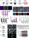
Characteristics of iPSC-derived astroglial progenitor cells and astrocytes. (A) Schematic representation of differentiation of healthy donor cells to astrocyte progenitors and astrocytes. (B) Representative immunographs and quantification of nestin and vimentin coexpression in APC cultures. (C) Representative immunographs of astrocytes of all lines immunostained for GFAP and GLAST from three replicate experiments. (D) A majority of cells of all lines are GFAP-expressing cells (iPSC1, 87.8 ± 1.7%; iPSC2, 91.4 ± 1.7%; and iPSC3, 89.2 ± 0.7%), with a minor proportion expressing the neuronal marker, NeuN (iPSC1, 6.3 ± 2%; iPSC2, 5.4 ± 1.8%; and iPSC3, 6 ± 0.4%). (E) Proliferating Ki67-positive cells in iPSC1, iPSC2, and iPSC3 APC and astrocyte cultures. (F) Functional evaluation of APC and astrocyte cultures by l-glutamate uptake assay. (G) Representative immunoblots of PrPC expression in MM (iPSC1), MV (iPSC2), and VV (iPSC3) lines as APC cultures and their corresponding CNTF differentiated astrocytes from two samples each in two replicate experiments. The amount of total protein loaded was 20 µg/lane. The blots were developed with anti-PrP 3F4 antibody and then stripped and reprobed with anti–β actin antibody as a loading control. Molecular masses are in kilodaltons. (H) Maximum intensity projection of Z stacks of VV (iPSC3) astrocytes immunolabeled for PrPC (green) and cell membrane (white) without and with Triton X-100 permeabilization. (I) Summary table of all cell lines and CJD strain combinations with their respective PRNP codon 129 polymorphisms used in this study. (J) Graphic representation of live/dead (viability/cytotoxicity) assay of iPSC1, iPSC2, and iPSC3 astrocytes exposed to 1% spin-filtered vCJD, sCJD, or NBHs for 24 h and analyzed immediately (0 dpe) or after 3 d of recovery in fresh media (3 dpe). Unexposed cells served as a baseline control for each cell line. Cell count of each group is represented as percentage of total cells, and the data were acquired from 10 randomized fields from three replicate experiments, unless otherwise stated. (B–F and J) Data are plotted as mean ± SEM and analyzed by one-way ANOVA, followed by Tukey’s multicolumn comparison test; *, P < 0.05; ****, P < 0.0001; ns, not significant. (B, C, and H) Nuclei were stained with DAPI (blue). Bars, 20 µm.
Figure 2.
Figure 2.
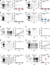
Human iPSC–derived astrocytes replicate PrPSc in vitro in a PRNP codon 129–dependent manner. APC and astrocyte cultures were analyzed by immunoblots immediately after 24 h of exposure (0 dpe), 3 and 8 dpe. (A) APC cultures of the MM (iPSC1) line exposed to 1% spin-filtered vCJD. APC cultures of the VV (iPSC3) line exposed to 1% spin-filtered vCJD (B), sCJD (VV1) (C), and sCJD (VV2) (D). (A–D) n = 1–3, in triplicate. (E) Astrocytes of the MM (iPSC1) genotype replicate PrPSc after exposure to 1% spin-filtered vCJD (MM) inoculum. (F) Astrocytes of MV (iPSC2) and (G) VV (iPSC3) genotypes exposed to 1% vCJD do not replicate PrPSc. (H) VV (iPSC3) astrocytes replicate PrPSc when exposed to 1% spin-filtered sCJD (VV2) brain homogenate. Representative immunoblots and linear regression of PK-resistant PrPSc level from n = 6 (E), n = 4 (F), n = 4 (G), and n = 4 (H) independent identical experiments generally performed in triplicate. (I) VV (iPSC3) astrocytes exposed to 1% spin-filtered sCJD (VV1) brain homogenate. (J) Independent MM (iPSC4) astrocytes exposed to 1% spin-filtered vCJD (MM). (I and J) n = 1–2, in triplicate. PK-resistant PrPSc signal values in cell lysates were normalized by the PrPSc signal value of the inoculum used in each individual experiment. A–D, I, and J are plotted including mean and analyzed by one-way ANOVA followed by Tukey’s multicolumn comparison test. (E–H) Mean ± SEM. Linear regression was applied to establish a trend line (black) that is shown with 95% confidence bands (black dotted). Dashed gray lines in immunoblot images (E, F, and J) indicate a montage image in which lanes of the same blot and exposure have been rearranged for presentation purposes. Note: the y axis in all cells exposed to vCJD (A, B, E–G, and J) has a fivefold-extended range because of high levels of vCJD PrPSc accumulation detected in MM (iPSC1 and iPSC4) astrocytes. Blots were developed using anti-PrP 3F4 (A, E, F, and J) and HuM-P (B–D and G–I) antibodies. Molecular mass is indicated in kilodaltons. *, P < 0.05; **, P < 0.01; ***, P < 0.001; ns, not significant.
Figure 3.
Figure 3.
Human iPSC–derived astrocytes accumulate PrPSc when the CJD inoculum and cell PRNP codon 129 genotype are matched. (A) PrPSc accumulation in MM (iPSC1) astrocytes was observed at 8 dpe to 1% spin-filtered vCJD inoculum (bright green). Quantification of PrP immunolabeling intensity (right) showed significant increase in cell-associated PrPSc when compared with PrPC of unexposed control cells. (B) MV (iPSC2) astrocytes fail to accumulate detectable PrPSc after exposure to 1% spin-filtered vCJD inoculum. Only the faint green immunolabeling of endogenous PrPC can be seen. (C) VV (iPSC3) astrocytes fail to accumulate PrPSc when exposed to 1% spin-filtered vCJD. (D) PrPSc accumulation (bright green) in (iPSC3) VV astrocytes exposed to 1% spin-filtered sCJD (VV2) inoculum and allowed to recover for 8 d. Quantification of PrP immunolabeling intensity (right) showed significant increase in cell-associated PrPSc at 8 dpe. Faint green endogenous PrPC signal is present in corresponding unexposed control cells in A–D. The data shown in A–D were generated from multiple fields from an experiment conducted in triplicate and are representative of experiments vCJD/iPSC1 (n = 3), vCJD/iPSC2 (n = 2), vCJD/iPSC3 (n = 4), and sCJD VV2/iPSC3 (n = 7). PrP signal was normalized by cell count. Data are plotted with mean ± SEM, analyzed by one-way ANOVA, and followed by Tukey’s multicolumn comparison test: ****, P < 0.0001; ns, not significant. Cells were immunolabeled with anti-PrP antibody 6H4 (A and B) and HuM-P (C and D). Nuclei were stained with DAPI (blue). Bars, 20 µm. ctrl, control.
Figure 4.
Figure 4.
Dose-dependent accumulation and subpassage of CJD prions in iPSC-derived astrocytes. (A) Immunoblot analysis of astrocytes exposed to vCJD (MM) brain homogenate at five concentrations for 24 h and assayed immediately (0 dpe) or after recovery in fresh media for 3 d (3 dpe). MM (iPSC1) astrocytes replicate PrPSc in a concentration-dependent manner. MV (iPSC2) and VV (iPSC3) astrocytes failed to replicate PrPSc at 3 dpe. The experiment was conducted twice with similar results. (B) MM (iPSC1) astrocytes exposed to 1% spin-filtered vCJD brain homogenate. Both first and second passage were analyzed at 8 dpe. In the second passage experiment, the naive MM (iPSC1) astrocytes were exposed to vCJD-infected cell homogenate diluted to match PrPSc level of original vCJD brain homogenate used for exposure of astrocytes in the first passage. n = 2, in duplicate. (C) VV (iPSC3) astrocytes exposed to 1% spin-filtered sCJD (VV2) brain homogenate (first passage) and sCJD VV2–infected astrocyte homogenate (second passage). The naive VV (iPSC3) astrocytes were exposed to a whole sCJD-infected cell homogenate (1:1), i.e., cell lysate of a single well served as an inoculum for a new well because of a lower efficiency of sCJD prion propagation. Both passages were analyzed at 8 dpe. n = 2, in triplicate. Blots were developed using anti-PrP (A and B) 3F4 and (C) HuM-P antibodies. Molecular mass is indicated in kilodaltons. (B and C) Data are plotted with mean. PK-resistant PrPSc signal values in cell lysates were normalized by the PrPSc signal value of the inoculum used in each individual experiment. (D) Representative PrP and GFAP immunolabeling in VV (iPSC3) astrocytes exposed to 1% spin-filtered sCJD (VV2) brain homogenate (first passage, middle). VV astrocytes exposed to spin-filtered cell homogenate of astrocytes propagating sCJD (VV2; second passage, right). Both were analyzed at 8 dpe. PrP immunolabeling intensity (bright green) showed an increase in cell-associated PrPSc when astrocytes of first sCJD passage were compared with PrPC of unexposed control cells (control, left), and PrPSc signal appeared more abundant in astrocytes in the second passage. n = 2, in triplicate. (E) Maximum intensity projection of Z stacks of VV (iPSC3) astrocytes immunolabeled for PrP at 8 dpe in cells exposed to sCJD (VV2) infected cell homogenate (right) and unexposed control (left). (D and E) Cells were immunolabeled with anti-PrP antibody 3F4. Nuclei were stained with DAPI (blue). Bars: (D) 20 µm; (E) 5 µm.
Figure 5.
Figure 5.
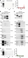
Differing kinetics of vCJD and sCJD prion propagation in human iPSC–derived astrocytes. In all experiments, astrocytes were exposed to 1% spin-filtered brain homogenate (24 h) and analyzed immediately (0 dpe) and at 8, 15, and 28 d later (8, 15, and 28 dpe). (A) Representative immunoblot of MV (iPSC2) astrocytes exposed to vCJD brain homogenate. Graphic representation of n = 2, duplicate and triplicate. (B) Immunoblots of MM (iPSC1) astrocytes exposed to 1% spin-filtered vCJD (lane 1), sCJD (MM1; lane 2), sCJD (VV2; lane 3), and NBH (lane 4). (C) Immunoblot of VV (iPSC3) astrocytes exposed to sCJD (MM1) brain homogenate. Representation of n = 2, in triplicate. (A and C) Data are plotted with mean. PK-resistant PrPSc signal values in cell lysates were normalized by the PrPSc signal value of the inoculum used in each individual experiment. (D) VV (iPSC3) astrocytes were exposed to vCJD (lane 1), sCJD (VV1; lane2), sCJD (VV2; lane 3), and NBH (lane 4). (B and D) Blots were developed at the same time/exposure; an example of n = 3 is shown. Blots were immunolabeled using anti-PrP 3F4 (A and B) and HuM-P (C and D) antibodies. Molecular mass is indicated in kilodaltons.

Comment in

  • A role for astroglia in prion diseases.
    Aguzzi A, Liu Y. Aguzzi A, et al. J Exp Med. 2017 Dec 4;214(12):3477-3479. doi: 10.1084/jem.20172045. Epub 2017 Nov 21. J Exp Med. 2017. PMID: 29162644 Free PMC article.

References

    1. Alibhai J., Blanco R.A., Barria M.A., Piccardo P., Caughey B., Perry V.H., Freeman T.C., and Manson J.C.. 2016. Distribution of misfolded prion protein seeding activity alone does not predict regions of neurodegeneration. PLoS Biol. 14:e1002579 10.1371/journal.pbio.1002579 - DOI - PMC - PubMed
    1. Arjona A., Simarro L., Islinger F., Nishida N., and Manuelidis L.. 2004. Two Creutzfeldt-Jakob disease agents reproduce prion protein-independent identities in cell cultures. Proc. Natl. Acad. Sci. USA. 101:8768–8773. 10.1073/pnas.0400158101 - DOI - PMC - PubMed
    1. Asuni A.A., Gray B., Bailey J., Skipp P., Perry V.H., and O’Connor V.. 2014. Analysis of the hippocampal proteome in ME7 prion disease reveals a predominant astrocytic signature and highlights the brain-restricted production of clusterin in chronic neurodegeneration. J. Biol. Chem. 289:4532–4545. 10.1074/jbc.M113.502690 - DOI - PMC - PubMed
    1. Berry D.B., Lu D., Geva M., Watts J.C., Bhardwaj S., Oehler A., Renslo A.R., DeArmond S.J., Prusiner S.B., and Giles K.. 2013. Drug resistance confounding prion therapeutics. Proc. Natl. Acad. Sci. USA. 110:E4160–E4169. 10.1073/pnas.1317164110 - DOI - PMC - PubMed
    1. Bishop M.T., Hart P., Aitchison L., Baybutt H.N., Plinston C., Thomson V., Tuzi N.L., Head M.W., Ironside J.W., Will R.G., and Manson J.C.. 2006. Predicting susceptibility and incubation time of human-to-human transmission of vCJD. Lancet Neurol. 5:393–398. 10.1016/S1474-4422(06)70413-6 - DOI - PubMed

Associated data