Review
. 2018 Feb;364(2):180-197.
doi: 10.1124/jpet.117.244673. Epub 2017 Nov 15.

Genetic and Molecular Regulation of Extrasynaptic GABA-A Receptors in the Brain: Therapeutic Insights for Epilepsy

Affiliations
Review

Genetic and Molecular Regulation of Extrasynaptic GABA-A Receptors in the Brain: Therapeutic Insights for Epilepsy

Shu-Hui Chuang et al. J Pharmacol Exp Ther. 2018 Feb.

Abstract

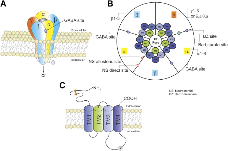
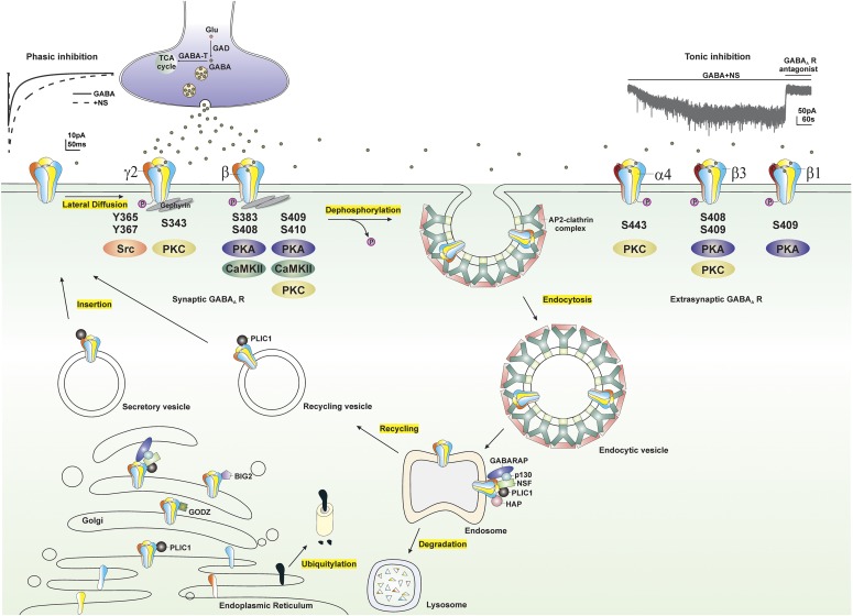
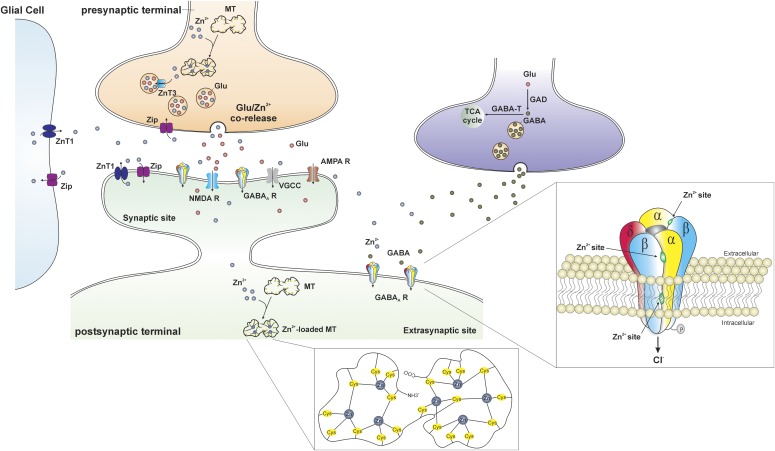
GABA-A receptors play a pivotal role in many brain diseases. Epilepsy is caused by acquired conditions and genetic defects in GABA receptor channels regulating neuronal excitability in the brain. The latter is referred to as GABA channelopathies. In the last two decades, major advances have been made in the genetics of epilepsy. The presence of specific GABAergic genetic abnormalities leading to some of the classic epileptic syndromes has been identified. Advances in molecular cloning and recombinant systems have helped characterize mutations in GABA-A receptor subunit genes in clinical neurology. GABA-A receptors are the prime targets for neurosteroids (NSs). However, GABA-A receptors are not static but undergo rapid changes in their number or composition in response to the neuroendocrine milieu. This review describes the recent advances in the genetic and neuroendocrine control of extrasynaptic and synaptic GABA-A receptors in epilepsy and its impact on neurologic conditions. It highlights the current knowledge of GABA genetics in epilepsy, with an emphasis on the neuroendocrine regulation of extrasynaptic GABA-A receptors in network excitability and seizure susceptibility. Recent advances in molecular regulation of extrasynaptic GABA-A receptor-mediated tonic inhibition are providing unique new therapeutic approaches for epilepsy, status epilepticus, and certain brain disorders. The discovery of an extrasynaptic molecular mechanism represents a milestone for developing novel therapies such as NS replacement therapy for catamenial epilepsy.


Figures

Fig. 1.
Fig. 1.
Schematic representation of typical GABA-A receptor (GABA-AR) structure and subunit composition. (A) GABA-ARs are heteropentamers forming a channel that is permeable to chloride ion passage. (B) A top view of the pentamer. GABA-ARs are made from a repertoire of 19 known subunits: α1–6, β1–3, γ1–3, δ, ε, θ, π, and ρ1–3. The most general stoichiometry of GABA-ARs contains two αs, two βs, and one γ; and the γ subunit can be substituted by δ, ε, θ, or π. Each subunit has four TMs (TM1–TM4). TM2s form a selective channel pore. GABA exerts fast inhibitory actions by activating postsynaptic GABA-ARs in the brain, causing the influx of negatively charged chloride ions and hyperpolarization of neurons, which serve to reduce neuronal excitability and firing. The GABA binding sites are located at the junction between subunit α and β, whereas benzodiazepines (BZs) bind at the interface between subunits α and γ. Barbiturates binding sites are distinct from the BZ binding site. The NSs have two putative binding sites including allosteric and direct binding sites. The allosteric binding site is located at the α subunit TMs, whereas the direct binding site is within the TMs of the α and β subunits. (C) GABA-ARs belong to the Cys-loop family of ligand-gated ion channels, which also contains nicotinic acetylcholine, glycine, and serotonin 5-HT3 receptors. Each subunit has one long extracellular N terminus that interacts with a variety of drugs including BZs, barbiturates, and NSs; four TMs (TM1–TM4); and one short intracellular loop that links TM1 and TM2, one short extracellular loop that links TM2 and TM3, one long intracellular loop that links TM3 and TM4 and can be modulated by phosphorylation, and one small extracellular C terminus.
Fig. 2.
Fig. 2.
GABA-A receptor subunit family. (A) A dendrogram illustrating the 19 known genes encoding for the mammalian GABA-A receptor subunits and the sequence homologies. (B) Chromosomal clusters of GABA-A receptor subunit genes.
Fig. 3.
Fig. 3.
Extrasynaptic GABA-A receptor δ subunit distribution. (A) The estimated abundance of GABA-A receptor subtypes in the rodent brain. (B) GABA-A receptor δ subunit distribution. *denotes δ subunit.
Fig. 4.
Fig. 4.
Summary outline of the association of genetic epilepsies with GABA-A receptor subunits and other genetic factors. (Right) GABA-A receptor subunits that are involved in genetic epilepsies. Mutations in each subunit are listed. (Left) Other genetic factors that are associated with genetic epilepsies. CACNA1H (Cav3.2), a voltage-dependent T-type calcium channel α1H subunit gene; CACNB4, a voltage-dependent L-type calcium channel β subunit gene; CPA6, a gene that encodes carboxypeptidase A6; CASR, a gene that encodes calcium sensor receptor; EFHC1, a gene that encodes a protein with an EF-hand domains; SCN1A, a sodium voltage-gated channel α1A subunit gene; SCN1B, a sodium voltage-gated channel α1B subunit gene; SCN2A, a sodium voltage-gated channel α2A subunit gene; SCN2B, a sodium voltage-gated channel α2B subunit gene.
Fig. 5.
Fig. 5.
Modulation of extrasynaptic GABA-A receptors (GABA-ARs) trafficking by phosphorylation. GABA-ARs composed of α, β, and γ subunits are mostly clustered at synaptic sites, whereas GABA-ARs composed of α, β, and δ subunits are clustered at extrasynaptic sites. GABA-ARs are assembled in the ER, matured in the Golgi, secreted, and inserted into the plasma membrane. Synaptic GABA-ARs, reaching their destination through lateral diffusion in the cell membrane, are activated by presynaptic release of GABA and produce fast and transient phasic inhibition, whereas continuous activation of extrasynaptic GABA-ARs by ambient GABA generates persistent tonic inhibition that sets the baseline of neuronal inhibition. NSs at submicromolar concentrations potentiate both phasic and tonic inhibition through allosteric modulation of synaptic and extrasynaptic GABA-ARs, respectively. GABA-AR subunits, including α4, β, and γ2 subunits, contain residues that can be phosphorylated by various protein kinases. Phosphorylation of residues within GABA-ARs not only regulates receptor function but maintains their surface expression. Dephosphorylation of these subunits triggers receptor internalization through adaptor protein 2 (AP2)-clathrin complex–dependent endocytosis. Internalized receptors are subsequently transported to the endosomal system, where they can be either recycled to the surface or degraded in the lysosomes. GABA-AR trafficking is facilitated by a variety of protein-protein interactions. BIG2, a brefeldin A-inhibited GDP/GTP exchange factor, binds to the intracellular loop of β subunits and is involved in the trafficking of receptors; GABARAP, a GABA-AR–associated protein, interacts with the γ2 subunit of GABA-ARs and might facilitate receptor insertion into the cell membrane; GABA-T, GABA transaminase; GODZ, a Golgi-specific DHHC zinc finger domain protein, mediates post-translational palmitoylation; HAP, a Huntingtin-associated protein, interacts with the β subunit of GABA-ARs and facilitates receptor recycling to the cell membrane; GAD, glutamate decarboxylase; NSF, a N-ethylmaleimide–sensitive factor, interacts with GABARAP and p130 and plays a crucial role in intracellular transport; p130, a phospholipase, shares the same binding site on the γ2 subunit with the GABARAP; PLIC-1, a ubiquitin-like protein, increases receptor stability and enhances membrane insertion of receptors.
Fig. 6.
Fig. 6.
Trafficking of Zn2+ at the gluzinergic synapse. In the gluzinergic terminal, the availability of free Zn2+ is regulated by metallothioneins (MTs), the primary intracellular Zn2+-buffering proteins. The MT is a dumbbell-shaped, cysteine (Cys)-rich protein composed of two domains in which 7 zinc atoms are tetrahedrally bound to 20 cysteines (inset, middle). The Zn2+ transporter (ZnT) and Zip proteins are also involved in the regulation of Zn2+ in the cytoplasm. ZnT proteins promote Zn2+ efflux and vesicular uptake to decrease the amount of intracellular Zn2+, whereas Zip proteins facilitate the influx of extracellular Zn2+ into neurons and glial cells to increase the concentration of intracellular Zn2+. Free Zn2+ is transferred into synaptic vesicles through the ZnT3 proteins and stored with glutamate. During normal neurotransmission, Zn2+- and glutamate-containing vesicles fuse with cell membrane and corelease Zn2+ and glutamate into the synaptic cleft. There are a variety of postsynaptic targets that Zn2+ can act on, including N-methyl-d-aspartate receptors (NMDA Rs), α-amino-3-hydroxy-5-methyl-4-isoxazolepropionic acid receptors (AMPA Rs), voltage-gated calcium channels (VGCCs), GABA-ARs, and a number of other channels, transporters, and receptors. Three distinct Zn2+ binding sites mediate its inhibition of extrasynaptic δ-containing GABA-ARs (inset, right): one site at the internal surface of the channel pore and two at the external amino terminus of the α-β interfaces.

References

    1. Abramian AM, Comenencia-Ortiz E, Modgil A, Vien TN, Nakamura Y, Moore YE, Maguire JL, Terunuma M, Davies PA, Moss SJ. (2014) Neurosteroids promote phosphorylation and membrane insertion of extrasynaptic GABA-A receptors. Proc Natl Acad Sci USA 111:7132–7137. - PMC - PubMed
    1. Abramian AM, Comenencia-Ortiz E, Vithlani M, Tretter EV, Sieghart W, Davies PA, Moss SJ. (2010) Protein kinase C phosphorylation regulates membrane insertion of GABAA receptor subtypes that mediate tonic inhibition. J Biol Chem 285:41795–41805. - PMC - PubMed
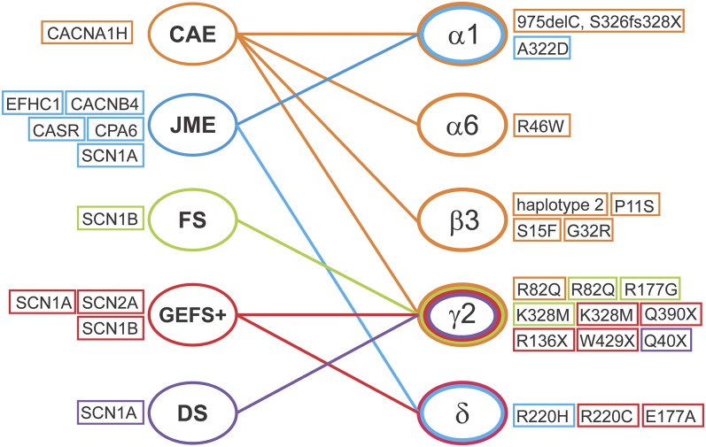
    1. Audenaert D, Schwartz E, Claeys KG, Claes L, Deprez L, Suls A, Van Dyck T, Lagae L, Van Broeckhoven C, Macdonald RL, et al. (2006) A novel GABRG2 mutation associated with febrile seizures. Neurology 67:687–690. - PubMed
    1. Barberis A, Cherubini E, Mozrzymas JW. (2000) Zinc inhibits miniature GABAergic currents by allosteric modulation of GABAA receptor gating. J Neurosci 20:8618–8627. - PMC - PubMed
    1. Baulac S, Huberfeld G, Gourfinkel-An I, Mitropoulou G, Beranger A, Prud’homme JF, Baulac M, Brice A, Bruzzone R, LeGuern E. (2001) First genetic evidence of GABA(A) receptor dysfunction in epilepsy: a mutation in the gamma2-subunit gene. Nat Genet 28:46–48. - PubMed

Publication types

Substances