. 2018 Jan 17;92(3):e01784-17.
doi: 10.1128/JVI.01784-17. Print 2018 Feb 1.

Interaction between a Unique Minor Protein and a Major Capsid Protein of Bluetongue Virus Controls Virus Infectivity

Affiliations

Interaction between a Unique Minor Protein and a Major Capsid Protein of Bluetongue Virus Controls Virus Infectivity

Eiko Matsuo et al. J Virol. .

Abstract

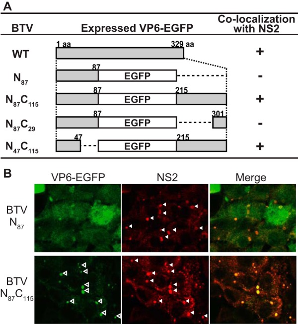
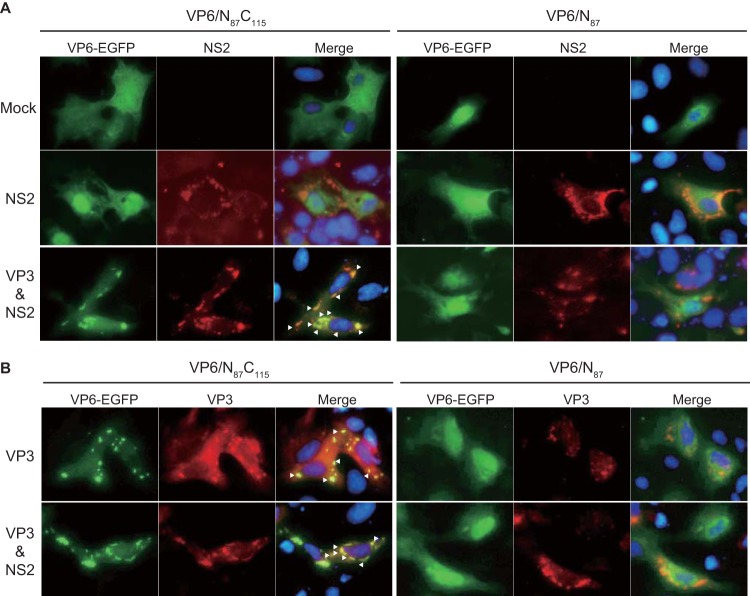
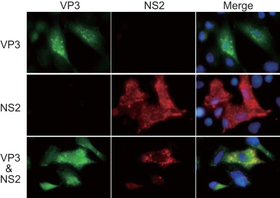
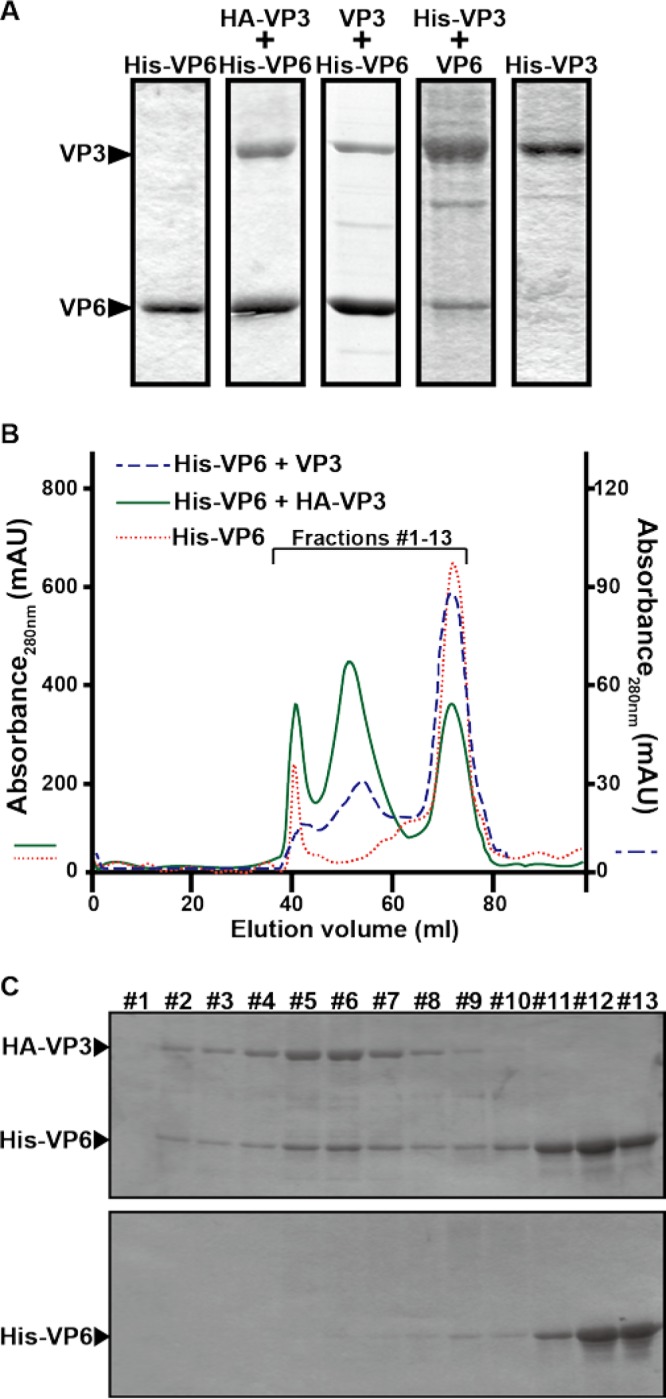
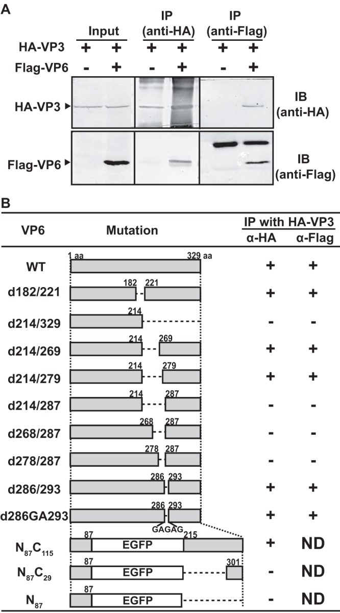
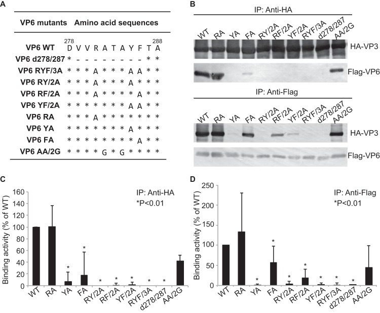
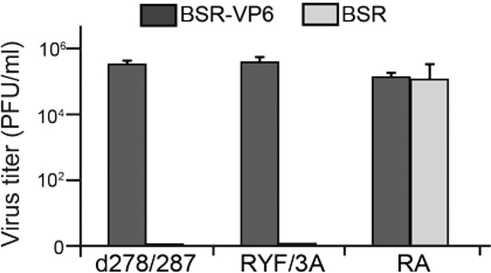
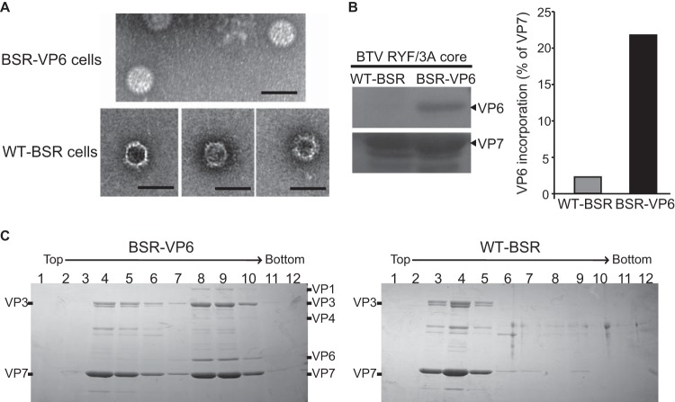
Among the Reoviridae family of double-stranded RNA viruses, only members of the Orbivirus genus possess a unique structural protein, termed VP6, within their particles. Bluetongue virus (BTV), an important livestock pathogen, is the prototype Orbivirus BTV VP6 is an ATP-dependent RNA helicase, and it is indispensable for virus replication. In the study described in this report, we investigated how VP6 might be recruited to the virus capsid and whether the BTV structural protein VP3, which forms the internal layer of the virus capsid core, is involved in VP6 recruitment. We first demonstrated that VP6 interacts with VP3 and colocalizes with VP3 during capsid assembly. A series of VP6 mutants was then generated, and in combination with immunoprecipitation and size exclusion chromatographic analyses, we demonstrated that VP6 directly interacts with VP3 via a specific region of the C-terminal portion of VP6. Finally, using our reverse genetics system, mutant VP6 proteins were introduced into the BTV genome and interactions between VP6 and VP3 were shown in a live cell system. We demonstrate that BTV strains possessing a mutant VP6 are replication deficient in wild-type BSR cells and fail to recruit the viral replicase complex into the virus particle core. Taken together, these data suggest that the interaction between VP3 and VP6 could be important in the packaging of the viral genome and early stages of particle formation.IMPORTANCE The orbivirus bluetongue virus (BTV) is the causative agent of bluetongue disease of livestock, often causing significant economic and agricultural impacts in the livestock industry. In the study described in this report, we identified the essential region and residues of the unique orbivirus capsid protein VP6 which are responsible for its interaction with other BTV proteins and its subsequent recruitment into the virus particle. The nature and mechanism of these interactions suggest that VP6 has a key role in packaging of the BTV genome into the virus particle. As such, this is a highly significant finding, as this new understanding of BTV assembly could be exploited to design novel vaccines and antivirals against bluetongue disease.

Keywords: BTV; VP6 translocation; reverse genetics system.


Figures

FIG 1
FIG 1
Localization of chimeric VP6-EGFP proteins in WT-BSR cells infected with various chimeric BTV strains. (A) Schematic representation of the changes introduced in BTV VP6. The name of the mutation is indicated on the left. Numbers in the middle column indicate amino acid (aa) positions in VP6, where EGFPs were fused with the N- and/or C-terminal region of VP6. +, colocalization of VP6-EGFP with NS2. (B) Colocalization of VP6 to which EGFP was fused at the C terminus with NS2. Either BTV/N87 (top) or BTV/N87C115 (bottom) was used to infect WT-BSR cells at an MOI of 1.0. At 24 h postinfection, the expression of EGFP and NS2 was observed using confocal microscopy. NS2 was detected using an anti-NS2 antibody produced in a guinea pig. Open and closed arrowheads, punctate structures of VP6 and VIBs, respectively.
FIG 2
FIG 2
Translocation of VP6 mutants in the presence of VP3. (A) Colocalization of VP6 to which EGFP was fused at the C terminus with NS2 in the presence of VP3. Each of the mammalian expression plasmids, pCAG-PBTV10-VP6/N87C115 (left) and pCAG-PBTV10-VP6/N87 (right), was cotransfected with either pCAG-PBTV1-NS2 alone (middle) or pCAG-PBTV1-NS2 with pCAG-PBTV1-VP3 (bottom) into WT-BSR cells. As a control, pCAG-PBTV10-VP6/N87C115 and pCAG-PBTV10-VP6/N87 was each transfected alone into the cells (top). At 24 h posttransfection, the distributions of VP6-EGFP were observed using fluorescence microscopy. NS2 was detected using a guinea pig anti-NS2 antibody. Arrowheads, colocalization of VP6 with NS2. Nuclei were detected using DAPI. (B) Colocalization of VP6 to which EGFP was fused at the C terminus with VP3. Either VP6/N87C115 (left) or VP6/N87 (right) was coexpressed with VP3, using mammalian expression plasmids, in the absence (top) or presence of NS2 (bottom) in WT-BSR cells. The distributions of VP6-EGFP were observed using fluorescence microscopy. VP3 was detected using a mouse anti-VP3 antibody. Nuclei were detected using DAPI. Arrowheads, colocalization of VP6-EGFP and VP3.
FIG 3
FIG 3
Colocalization of VP3 with NS2. VP3 was coexpressed with NS2 in WT-BSR cells (bottom). As a control, either VP3 (top) or NS2 (middle) was singly expressed in the cells. VP3 and NS2 were detected using a mouse anti-VP3 antibody and a guinea pig anti-NS2 antibody, respectively.
FIG 4
FIG 4
Interaction of VP6 with VP3. (A) Copurification of VP6 with VP3, each of which was expressed using a baculovirus expression system. His-tagged VP6 (His-VP6) was coexpressed with either HA-tagged VP3 (HA-VP3) or nontagged VP3 (VP3) in Sf9 cells. In parallel, His-tagged VP3 (VP3) was coexpressed with nontagged VP6 (VP6). As a control, either His-VP6 or His-VP3 was expressed. Proteins were purified with a His-Select nickel affinity gel. (B) Analysis of the VP6/VP3 complex by gel filtration chromatography. Two types of VP3/VP6 complexes, His-VP6/HA-VP3 (green solid line) and His-VP6/VP3 (blue dashed line), copurified using nickel affinity gels, were loaded onto an equilibrated HiPrep 16/60 Sephacryl S-300 HR gel filtration column and eluted with the same buffer at a flow rate of 0.5 ml/min. His-VP6 (red dotted line) was loaded as a control. The left vertical axis indicates the absorbance at 280 nm of His-VP6 and the His-VP6/HA-VP3 complex. The right vertical axis indicates the absorbance at 280 nm of the His-VP6/VP3 complex. mAU, milli-absorbance units. (C) Fractions of the His-VP6/HA-VP3 complex (fractions 1 to 13) were collected between elution volumes of 38 ml and 77 ml and analyzed using SDS-PAGE (top). As a control, the same fractions of His-VP6 were analyzed (bottom).
FIG 5
FIG 5
Direct interaction of VP6 with VP3 at a region between residues 279 and 286. (A) Coimmunoprecipitation of VP6 with VP3. HA-tagged VP3 (HA-VP3) was expressed in WT-BSR cells in the presence or absence of Flag-tagged VP6 (Flag-VP6). HA-VP3 and Flag-VP6 were immunoprecipitated using a mouse anti-HA MAb and a mouse anti-Flag MAb, respectively. Precipitated proteins were detected by immunoblotting using a rabbit anti-HA pAb and a rabbit anti-Flag pAb, respectively. (B) Schematic representation of modified VP6. Numbers in the middle column indicate amino acid positions in VP6 where changes were introduced. Note that no Flag tag was inserted at the N-terminal end of the three VP6 mutants N87C115, N87C29, and N87. +, coimmunoprecipitation; IP, immunoprecipitation; IB, immunoblotting; aa, amino acid; ND, not determined.
FIG 6
FIG 6
Mapping of the amino acid residues of VP6 essential for VP3 binding. (A) Schematic representation of the changes introduced into Flag-VP6. The name of the mutation is indicated on the left. Numbers indicate amino acid positions according to the VP6 amino acid sequence. The amino acid changes are also shown. Asterisks indicate no change, and dashes indicate deletions. (B) The interaction between HA-VP3 and a series of Flag-VP6 mutants was analyzed by immunoprecipitation using either anti-HA MAb (top) or anti-Flag MAb (bottom). (C) The relative densities of Flag-VP6 mutant binding to HA-VP3 were quantified using gray-value analysis and normalized by the relative densities of HA-VP3. The activity of binding of each Flag-VP6 mutant to HA-VP3 was calculated in five experiments, and the results (means ± SDs) are shown as a percentage of the binding of the WT. *, a significant difference in comparison to the binding activity of WT Flag-VP6 (P < 0.01). (D) The relative density of HA-VP3 binding to each of the Flag-VP6 mutants was quantified using gray-value analysis and normalized by the relative density of each Flag-VP6 mutant. The activity of HA-VP3 binding to each Flag-VP6 mutant was calculated in five experiments, and the results (means ± SDs) are shown as a percentage of the binding of the WT. *, a significant difference in comparison to the activity of HA-VP3 binding to WT Flag-VP6 (P < 0.01).
FIG 7
FIG 7
Assay of VP6/VP3 interaction-defective BTV, BTV RYF/3A (RYF/3A), and BTV d278/287 (d278/287) replication. Each 100 μl (∼1 × 10³ PFU) of VP6/VP3 interaction-defective BTV once amplified in BSR-VP6 cells was inoculated into either WT-BSR cells (light gray) or BSR-VP6 cells (dark gray). At 24 h postinoculation, the total virus titer (mean ± SD) was determined by plaque assay. As a control, cells were infected with BTV RA (RA).
FIG 8
FIG 8
Localization of VP6, VP3, and NS2 in BTV-infected cells. WT-BSR cells were infected with each of WT-BTV (A), RA-BTV (B), RYF/3A-BTV (C), and d278/287-BTV (D), expressing WT VP6, RA VP6, RYF/3A VP6, and d278/287 VP6, respectively. Note that the cells were infected with WT-BTV and RA-BTV at an MOI of 0.1. At 24 h postinfection, the expression of VP6, VP3, and NS2 was observed using confocal microscopy. VP6 was detected using a guinea pig anti-VP6 antibody (top) and a rabbit anti-VP6 antibody (middle). VP3 was detected using a mouse anti-VP3 antibody. NS2 was detected using a guinea pig anti-NS2 antibody.
FIG 9
FIG 9
Nonrecruitment of VP6/VP3 interaction-defective VP6 as well as VP1, VP4, and RNAs into core particles purified from WT-BSR cells. (A) Electron microscopy of BTV RYF/3A core particles amplified in either BSR-VP6 cells (top) or normal BSR cells (bottom). Bars, 100 nm. (B) The incorporation of VP6 proteins with core particles was analyzed using immunoblotting (left). VP6 proteins were normalized with VP7, and the level of incorporation with VP6 into the particles is shown as a percentage of that for VP7 proteins (right). (C) Analysis of VP6/VP3 interaction-defective BTV core particles purified from WT-BSR and BSR-VP6 cells by CsCl gradient centrifugation. Supernatants from BSR-VP6 (left) or BSR (right) infected cell lysates were spun down over a 30% (wt/vol) sucrose cushion and subjected to CsCl equilibrium centrifugation. Thirteen fractions were collected from the top, and then 12 fractions from the top were analyzed by SDS-PAGE. The gels were stained with Coomassie brilliant blue.

References

    1. Roy P. 2013. Orbiviruses, p 1402–1423. In Knipe DM, Howley PM, Cohen JI, Griffin DE, Lamb RA, Martin MA, Racaniello VR, Roizman B (ed), Fields virology, 6th ed, vol 2 Lippincott Williams & Wilkins, Philadelphia, PA.
    1. Matsuo E, Celma CC, Boyce M, Viarouge C, Sailleau C, Dubois E, Breard E, Thiery R, Zientara S, Roy P. 2011. Generation of replication-defective virus-based vaccines that confer full protection in sheep against virulent bluetongue virus challenge. J Virol 85:10213–10221. doi:10.1128/JVI.05412-11. - DOI - PMC - PubMed
    1. Matsuo E, Roy P. 2009. Bluetongue virus VP6 acts early in the replication cycle and can form the basis of chimeric virus formation. J Virol 83:8842–8848. doi:10.1128/JVI.00465-09. - DOI - PMC - PubMed
    1. Matsuo E, Roy P. 2013. Minimum requirements for bluetongue virus primary replication in vivo. J Virol 87:882–889. doi:10.1128/JVI.02363-12. - DOI - PMC - PubMed
    1. Stauber N, Martinez-Costas J, Sutton G, Monastyrskaya K, Roy P. 1997. Bluetongue virus VP6 protein binds ATP and exhibits an RNA-dependent ATPase function and a helicase activity that catalyze the unwinding of double-stranded RNA substrates. J Virol 71:7220–7226. - PMC - PubMed

Publication types