. 2018 Jan 17;92(3):e01933-17.
doi: 10.1128/JVI.01933-17. Print 2018 Feb 1.

Entry of Human Coronavirus NL63 into the Cell

Affiliations

Entry of Human Coronavirus NL63 into the Cell

Aleksandra Milewska et al. J Virol. .

Abstract

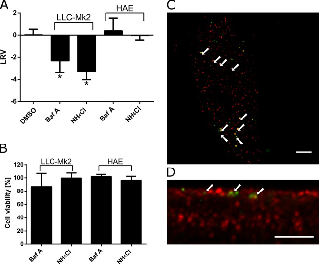
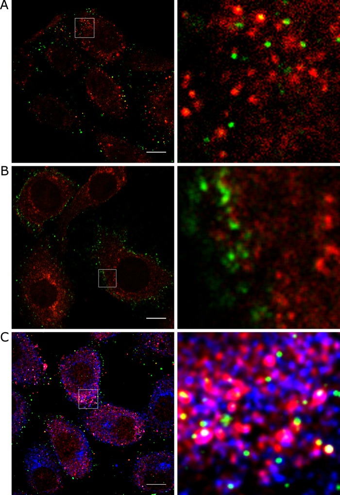
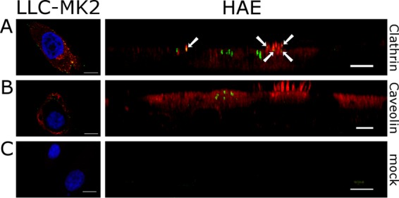
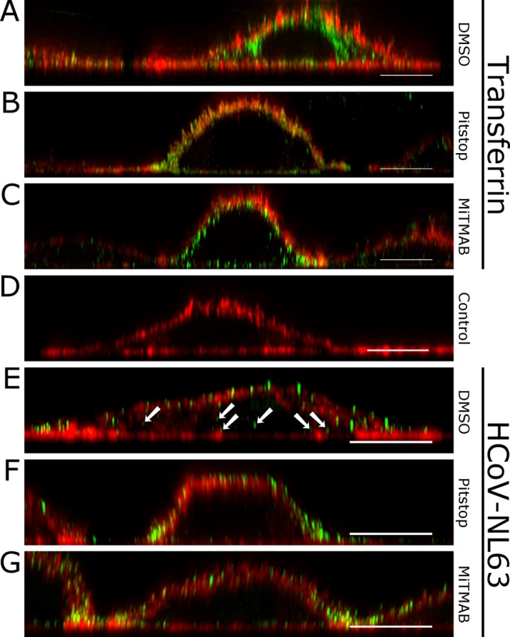
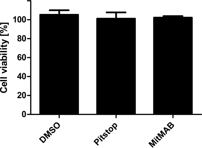
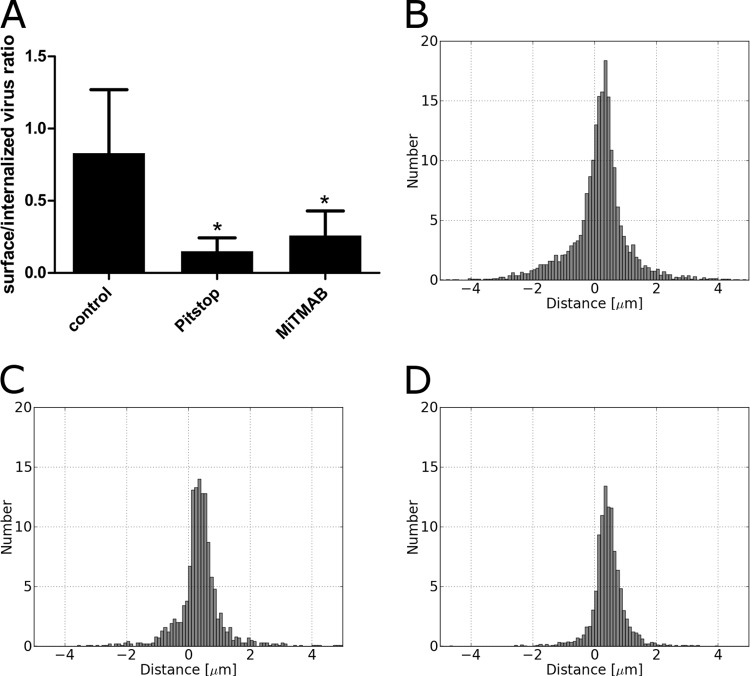
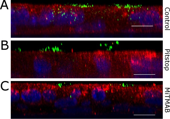
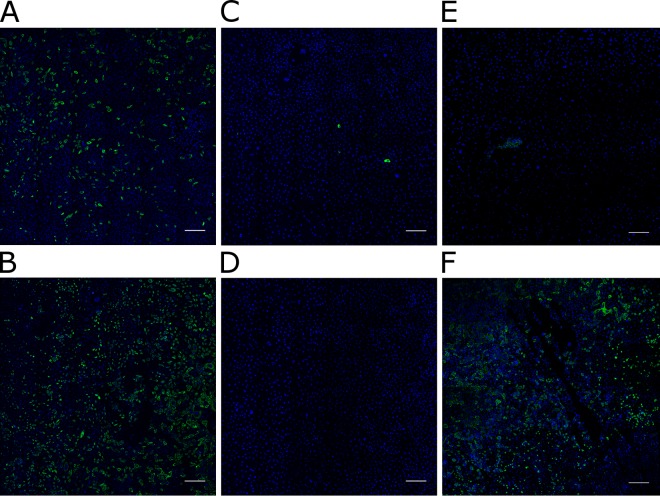
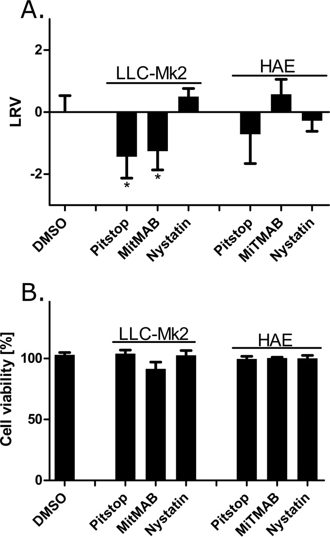
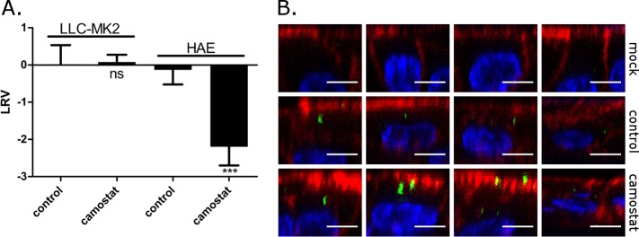
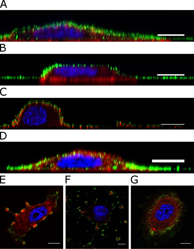
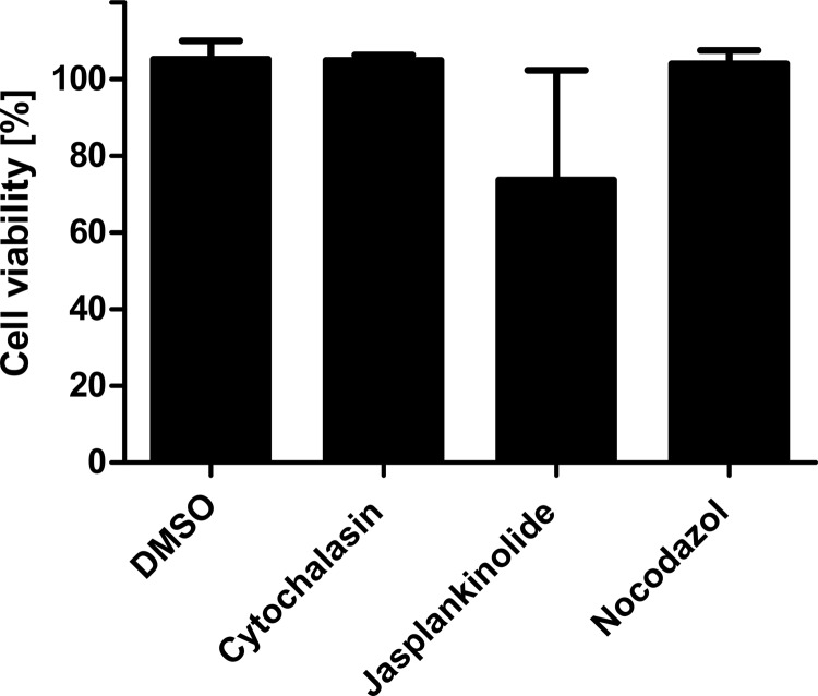
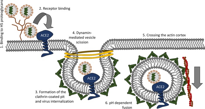
The first steps of human coronavirus NL63 (HCoV-NL63) infection were previously described. The virus binds to target cells by use of heparan sulfate proteoglycans and interacts with the ACE2 protein. Subsequent events, including virus internalization and trafficking, remain to be elucidated. In this study, we mapped the process of HCoV-NL63 entry into the LLC-Mk2 cell line and ex vivo three-dimensional (3D) tracheobronchial tissue. Using a variety of techniques, we have shown that HCoV-NL63 virions require endocytosis for successful entry into the LLC-MK2 cells, and interaction between the virus and the ACE2 molecule triggers recruitment of clathrin. Subsequent vesicle scission by dynamin results in virus internalization, and the newly formed vesicle passes the actin cortex, which requires active cytoskeleton rearrangement. Finally, acidification of the endosomal microenvironment is required for successful fusion and release of the viral genome into the cytoplasm. For 3D tracheobronchial tissue cultures, we also observed that the virus enters the cell by clathrin-mediated endocytosis, but we obtained results suggesting that this pathway may be bypassed.IMPORTANCE Available data on coronavirus entry frequently originate from studies employing immortalized cell lines or undifferentiated cells. Here, using the most advanced 3D tissue culture system mimicking the epithelium of conductive airways, we systematically mapped HCoV-NL63 entry into susceptible cells. The data obtained allow for a better understanding of the infection process and may support development of novel treatment strategies.

Keywords: Coronaviridae; HCoV-NL63; clathrin; coronavirus; endocytosis; entry; infection; internalization.


Figures

FIG 1
FIG 1
Importance of endosomal entry for HCoV-NL63 infection. (A) Inhibition of HCoV-NL63 infection in LLC-Mk2 cells and HAE cultures by the lysosomotropic agents ammonium chloride (NH4Cl) (50 mM) and bafilomycin A (Baf A) (100 nM), as determined by RT-qPCR. Data on the y axis represent LRVs. The assay was performed in triplicate, and average values with standard errors are presented. P values of <0.05 were considered significant and are denoted with an asterisk. (B) The cytotoxicity of the tested inhibitors was measured with an XTT assay. Data on the y axis represent viability of the treated cells compared to the untreated reference samples. The assay was performed in triplicate, and average values with standard errors are presented. (C and D) Confocal images showing colocalization of HCoV-NL63 virions with the early endosomal marker EEA1 on LLC-Mk2 cells (C) and HAE cultures (D). Scale bars = 5 μm. Green, HCoV-NL63; red, EEA1.
FIG 2
FIG 2
HCoV-NL63 binding to ACE2 triggers clathrin-mediated endocytosis. Precooled LLC-Mk2 cells were incubated with gradient-purified HCoV-NL63 for 40 min at 4°C following 0 min (A and B) or 5 min (C) of incubation at 32°C. Colocalization of the virus (green) and ACE2 (red) was analyzed using confocal microscopy (A). No colocalization with clathrin was observed after 0 min of incubation (B). Triple colocalization of virus with ACE2 and clathrin (blue) is visible in panel C. Images on the right side are zoomed-in regions indicated by white rectangles on the left-side slides. A representative image is shown. Scale bars = 10 μm.
FIG 3
FIG 3
HCoV-NL63 colocalizes with clathrin but not caveolin. (A and B) LLC-Mk2 cells were incubated with gradient-purified HCoV-NL63 for 40 min at 4°C following 5 min (A) or 20 min (B) of incubation at 32°C. HAE cultures were incubated with gradient-purified HCoV-NL63 for 40 min at 4°C following 120 min of incubation at 32°C. HCoV-NL63 colocalization with clathrin (A) or caveolin (B) was analyzed with confocal microscopy (HCoV-NL63, green; clathrin and caveolin, red; nuclei, blue). (C) Cells mock incubated and stained with isotypic antibodies were used as a control. Scale bars = 5 μm.
FIG 4
FIG 4
Clathrin and dynamin inhibitors hamper internalization of HCoV-NL63. (A to C) In order to verify the effectiveness of inhibitors, LLC-Mk2 cells were incubated with control DMSO (A), 10 μM Pitstop 2 (B), or 10 μM MiTMAB (C) for 30 min at 37°C and inoculated with Alexa Fluor 488-labeled transferrin. Following incubation (45 min, 37°C), cells were fixed and stained for actin (red). Transferrin entry was evaluated with confocal microscopy. (E to G) LLC-Mk2 cells were incubated with control DMSO (E), 10 μM Pitstop 2 (F), or 10 μM MiTMAB (G) for 30 min at 37°C. Cells were inoculated with purified HCoV-NL63 and incubated at 32°C for 1 h. Subsequently, cells were fixed and immunostained for HCoV-NL63 particles (green) and actin (red). (D) Mock-infected cells were used as a control. Scale bars = 10 μm.
FIG 5
FIG 5
Cytotoxicity of Pitstop 2 and MiTMAB on LLC-Mk2 cells. The cytotoxicity of the endocytosis inhibitors was tested with an XTT assay. Cells were incubated with control DMSO, 10 μM Pitstop 2, or 10 μM MiTMAB for 2 h at 37°C Data on the y axis represent viability of the treated cells compared to the untreated reference samples. The assay was performed in triplicate, and average values with standard errors are presented.
FIG 6
FIG 6
Numerical image analysis: clathrin and dynamin inhibitors block HCoV-NL63 entry. (B to D) LLC-Mk2 cells were incubated with DMSO (B), 10 μM MiTMAB (C), or 10 μM Pitstop 2 (D) for 30 min at 37°C and subsequently inoculated with purified HCoV-NL63 and incubated for 45 min at 32°C. Confocal images were digitalized, and the localization of each virus particle relative to the cellular membrane was assessed. (A) Number of internalized virus particles relative to number of virions on the cell surface (y axis) for cells treated with DMSO (control), Pitstop 2, or MiTMAB. In panels B, C, and D, raw data for cells treated with DMSO, Pitstop 2, or MiTMAB, respectively, are presented. Histograms show the average number of virus particles (y axis) versus the distance from the cell surface (x axis). Values of <0 on the x axis indicate that the virus is inside the cell, while for extracellular virions, the x value is ≥0.
FIG 7
FIG 7
Clathrin and dynamin inhibitors prevent HCoV-NL63 from entering the cell in the HAE model. HAE cultures were incubated with control DMSO (A), 10 μM Pitstop 2 (B), or 10 μM MiTMAB (C) for 1 h at 37°C. Cells were then inoculated with purified HCoV-NL63 and incubated at 32°C for 2 h. Subsequently, cells were fixed and immunostained for HCoV-NL63 particles (green), actin (red), and nuclei (blue). Scale bars = 5 μm.
FIG 8
FIG 8
Cytotoxicity of Pitstop 2 and MiTMAB on HAE cultures. The cytotoxicity of the endocytosis inhibitors was tested with an XTT assay. Cells were incubated with control DMSO, 10 μM Pitstop 2, or 10 μM MiTMAB for 2 h at 37°C. Data on the y axis represent the viability of the treated cells compared to the untreated reference samples. The assay was performed in triplicate, and average values with standard errors are presented.
FIG 9
FIG 9
Clathrin and dynamin inhibitors limit the number of LLC-Mk2 infected cells. (A to D) LLC-Mk2 cells were incubated with control DMSO (A), 5 μg/ml nystatin (B), 10 μM MiTMAB (C), or 10 μM Pitstop 2 (D) for 1 h at 37°C and inoculated with HCoV-NL63 (TCID50 = 100/ml). After 2 h of incubation at 32°C, virions that were not internalized were inactivated with acidic buffer (pH 3), and cells were incubated for 4 days at 32°C in the presence of the tested inhibitors or control DMSO. (E and F) The identical procedure was applied to cells, but in these MiTMAB (E) and Pitstop 2 (F) were applied after the acid wash. Fixed cells were immunostained with anti-NL63 nucleocapsid protein (green) and nuclei (blue), and confocal images were collected. Scale bar = 200 μm.
FIG 10
FIG 10
Clathrin and dynamin inhibitors hamper replication of HCoV-NL63 in LLC-MK2 cells. (A) HCoV-NL63 replication in LLC-Mk2 cells and HAE cultures in the presence of entry inhibitors or control DMSO was analyzed using RT-qPCR. Cultures were incubated with 10 μM Pitstop 2, 10 μM MiTMAB, 5 μg/ml nystatin, or DMSO for 1 h at 37°C and inoculated with HCoV-NL63 (TCID50 = 400/ml). After 2 h of incubation at 32°C, virions that were not internalized were inactivated with acidic buffer (pH 3), and cells were incubated for 5 days at 32°C. The data are presented as log reduction value (LRV) compared to the control sample. The assay was performed in triplicate, and average values with standard errors are presented. P values of <0.05 were considered significant and are denoted with an asterisk. (B) The cytotoxicity of the inhibitors was tested with an XTT assay. Cells were incubated with 10 μM Pitstop 2, 10 μM MiTMAB, 5 μg/ml nystatin, or DMSO for 5 days at 32°C. Data on the y axis represent viability of the treated cells compared to the untreated reference samples. The assay was performed in triplicate, and average values with standard errors are presented.
FIG 11
FIG 11
TMPRSS2 is required for entry into HAE cells but does not enable virus-cell fusion on the cell surface. (A) HCoV-NL63 replication in LLC-Mk2 cells and HAE cultures in the presence of camostat or control DMSO was analyzed using RT-qPCR. Cultures were incubated with 100 μM camostat or DMSO for 1 h at 37°C and inoculated with HCoV-NL63 (TCID50 = 400/ml). After 2 h of incubation at 32°C, virions that were not internalized were inactivated with acidic buffer (pH 3), and cells were incubated for 5 days at 32°C. The data are presented as log reduction value (LRV) compared to the control sample. The assay was performed in triplicate, and average values with standard errors are presented. P values of <0.05 were considered significant and are denoted with asterisks. (B) HAE cultures were incubated with control DMSO or 100 μM camostat for 1 h at 37°C. Further, cells were inoculated with purified HCoV-NL63 and incubated at 32°C for 2 h. Subsequently, cells were fixed and immunostained for HCoV-NL63 particles (green), actin (red), and nuclei (blue). Scale bars = 5 μm.
FIG 12
FIG 12
Actin is important for HCoV-NL63 entry. LLC-MK2 cells were incubated with DMSO (A), 10 μM cytochalasin D (B and E), 1.5 μM jasplakinolide (C and F), or 400 nM nocodazole (D and G) for 1 h at 37°C and then inoculated with purified HCoV-NL63 and incubated at 32°C for 1 h. Actin and virus localization was verified with confocal microscopy; fixed cells were immunostained for HCoV-NL63 particles (green), actin (red), and nuclei (blue). Scale bars = 10 μm.
FIG 13
FIG 13
Cytotoxicity of the cytoskeleton-modifying compounds. The cytotoxicity of the endocytosis inhibitors was tested with an XTT assay. Cells were incubated with DMSO, 10 μM cytochalasin D, 1.5 μM jasplakinolide, or 400 nM nocodazole for 2 h at 37°C. Data on the y axis represent viability of the treated cells compared to the untreated reference samples. The assay was performed in triplicate, and average values with standard errors are presented.
FIG 14
FIG 14
Early events during HCoV-NL63 infection.

References

    1. van der Hoek L, Pyrc K, Jebbink MF, Vermeulen-Oost W, Berkhout RJ, Wolthers KC, Wertheim-van Dillen PM, Kaandorp J, Spaargaren J, Berkhout B. 2004. Identification of a new human coronavirus. Nat Med 10:368–373. doi:10.1038/nm1024. - DOI - PMC - PubMed
    1. Konca C, Korukluoglu G, Tekin M, Almis H, Bucak İ Uygun H, Altas AB, Bayrakdar F. 2017. The first infant death associated with human Coronavirus NL63 infection. Pediatr Infect Dis J 36:231–233. doi:10.1097/INF.0000000000001390. - DOI - PubMed
    1. Mayer K, Nellessen C, Hahn-Ast C, Schumacher M, Pietzonka S, Eis-Hübinger AM, Drosten C, Brossart P, Wolf D. 2016. Fatal outcome of human coronavirus NL63 infection despite successful viral elimination by IFN-alpha in a patient with newly diagnosed ALL. Eur J Haematol 97:208–210. doi:10.1111/ejh.12744. - DOI - PMC - PubMed
    1. Cabeça TK, Bellei N. 2012. Human coronavirus NL-63 infection in a Brazilian patient suspected of H1N1 2009 influenza infection: description of a fatal case. J Clin Virol 53:82–84. doi:10.1016/j.jcv.2011.09.006. - DOI - PMC - PubMed
    1. Oosterhof L, Christensen CB, Sengeløv H. 2010. Fatal lower respiratory tract disease with human coronavirus NL63 in an adult haematopoietic cell transplant recipient. Bone Marrow Transplant 45:1115–1116. doi:10.1038/bmt.2009.292. - DOI - PMC - PubMed

Publication types

MeSH terms