. 2018 May;89(5):461-466.
doi: 10.1136/jnnp-2017-316853. Epub 2017 Nov 15.

Imaging and CSF analyses effectively distinguish CJD from its mimics

Affiliations

Imaging and CSF analyses effectively distinguish CJD from its mimics

Peter Rudge et al. J Neurol Neurosurg Psychiatry. 2018 May.

Abstract

Objective: To review clinical and investigation findings in patients referred to a specialist prion clinic who were suspected to have sporadic Creutzfeldt-Jakob disease (sCJD) and yet were found to have an alternative final diagnosis.

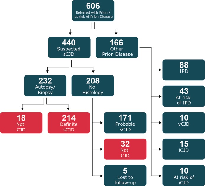
Methods: Review the clinical findings and investigations in 214 patients enrolled into the UK National Prion Monitoring Cohort Study between October 2008 and November 2015 who had postmortem confirmed sCJD and compare these features with 50 patients referred over the same period who had an alternative final diagnosis (CJD mimics).

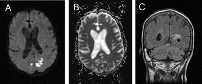
Results: Patients with an alternative diagnosis and those with sCJD were of similar age, sex and frequency of dementia but CJD mimics had a longer clinical history. Myoclonus, rigidity and hallucinations were more frequent in patients with sCJD but these features were not helpful in classifying individual patients. Alzheimer's disease, dementia with Lewy bodies and genetic neurodegenerative disorders were alternative diagnoses in more than half of the CJD mimic cases, and 10% had an immune-mediated encephalopathy; lymphoma, hepatic encephalopathy and progressive multifocal leukoencephalopathy were seen more than once. Diffusion-weighted MRI was the most useful readily available test to classify cases correctly (92% CJD, 2% CJD mimics). The CSF cell count, 14-3-3 protein detection and S100B were of limited value. A positive CSF RT-QuIC test, introduced during the course of the study, was found in 89% of tested CJD cases and 0% CJD mimics.

Conclusion: The combination of diffusion-weighted MRI analysis and CSF RT-QuIC allowed a perfect classification of sCJD versus its mimics in this study.


Conflict of interest statement

Competing interests: JC is a director and shareholder of D-Gen Ltd (London), an academic spin-out company working in the field of prion disease diagnosis, decontamination and therapeutics.

Figures

Figure 1
Figure 1
Flow chart of recruitment of all cases suspected of prion disease referred to the National Prion Clinic from 2008 to 2015. Patients included in the current study shown in red. sCJD, sporadic Creutzfeldt-Jakob disease.
Figure 2
Figure 2
Different MRI sequences in patients with definite sCJD showing the value of DWI confirmed on ADC measurement compared to FLAIR. Images A–C illustrate typical basal ganglia abnormalities on FLAIR (A), DWI (B) and ADC (C). Images E and F show typical features of ‘cortical ribboning’ in the frontal and parietal cortex on FLAIR (D), DWI (E) and ADC (F) sequences. ADC, apparent diffusion coefficient; DWI, diffusion-weighted images; sCJD, sporadic Creutzfeldt-Jakob disease.
Figure 3
Figure 3
MRI in patient with cerebral lymphoma. Note patchy restricted diffusion in the posterior cortex (A) with partial confirmation on ADC (B) together with abnormal signal in the periventricular area and white matter on FLAIR (C). These areas corresponded to leptomeningeal nodular enhancing soft tissue. ADC, apparent diffusion coefficient.
Figure 4
Figure 4
MRI in patient with autoimmune encephalopathy due to LGI1 antibodies. Note the high signal return from the swollen left basal ganglia on T2 (A), FLAIR (B) and DWI (C) sequences but restriction is not confirmed on ADC mapping (D). ADC, apparent diffusion coefficient; DWI, diffusion-weighted images; LGI1, Leucine-rich, glioma-inactivated antibodies-1.
Figure 5
Figure 5
MRI in a patient with autoimmune encephalopathy due to NMDA antibodies is similar to the scans seen in vCJD. Note pulvinar sign on FLAIR (A) and DWI (B). The ADC scan did not confirm restriction (C). There is minor periventricular high signal in the FLAIR sequence (A). ADC, apparent diffusion coefficient; DWI, diffusion-weighted images; NMDA, N-methyl-D-aspartate antibodies.

References

    1. Murray K. Creutzfeldt-Jacob disease mimics, or how to sort out the subacute encephalopathy patient. Pract Neurol 2011;11:19–28. 10.1136/jnnp.2010.235721 - DOI - PubMed
    1. Geschwind MD, Shu H, Haman A, et al. Rapidly progressive dementia. Ann Neurol 2008;64:97–108. 10.1002/ana.21430 - DOI - PMC - PubMed
    1. Zerr I, Kallenberg K, Summers DM, et al. Updated clinical diagnostic criteria for sporadic Creutzfeldt-Jakob disease. Brain 2009;132:2659–68. 10.1093/brain/awp191 - DOI - PMC - PubMed
    1. Hyare H, Thornton J, Stevens J, et al. High-b-value diffusion MR imaging and basal nuclei apparent diffusion coefficient measurements in variant and sporadic Creutzfeldt-Jakob disease. AJNR Am J Neuroradiol 2010;31:521–6. 10.3174/ajnr.A1860 - DOI - PMC - PubMed
    1. Carswell C, Thompson A, Lukic A, et al. MRI findings are often missed in the diagnosis of Creutzfeldt-Jakob disease. BMC Neurol 2012;12:153 10.1186/1471-2377-12-153 - DOI - PMC - PubMed

Publication types

Supplementary concepts