. 2017 Nov 16;8(1):1531.
doi: 10.1038/s41467-017-01494-4.

Super-achromatic monolithic microprobe for ultrahigh-resolution endoscopic optical coherence tomography at 800 nm

Affiliations

Super-achromatic monolithic microprobe for ultrahigh-resolution endoscopic optical coherence tomography at 800 nm

Wu Yuan et al. Nat Commun. .

Erratum in

Abstract

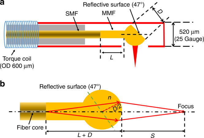
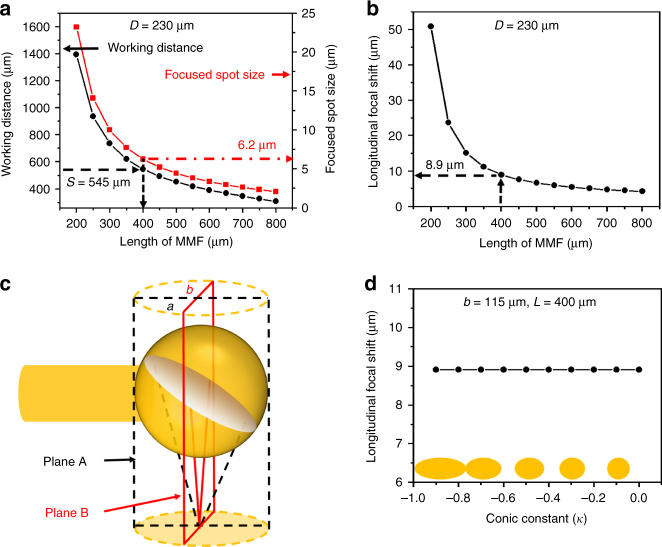
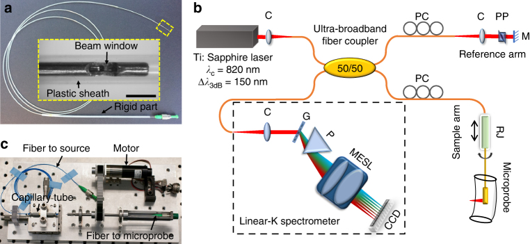
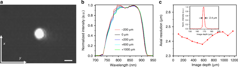
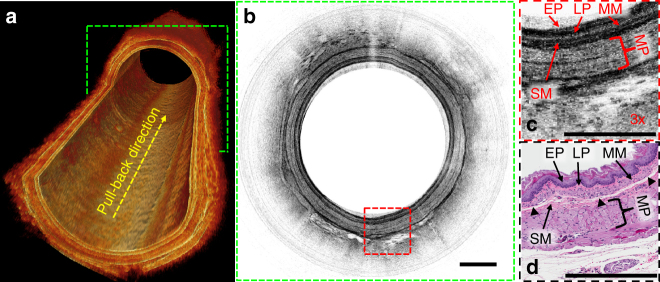
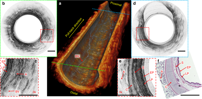
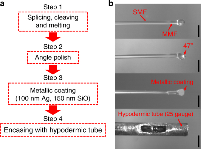
Endoscopic optical coherence tomography (OCT) has emerged as a valuable tool for advancing our understanding of the histomorphology of various internal luminal organs and studying the pathogenesis of relevant diseases. To date, this technology affords limited resolving power for discerning subtle pathological changes associated with early diseases. In addition, it remains challenging to access small luminal organs or pass through narrow luminal sections without potentially causing trauma to tissue with a traditional OCT endoscope of a 1-1.5 mm diameter. Here we report an ultracompact (520 µm in outer diameter and 5 mm in rigid length) and super-achromatic microprobe made with a built-in monolithic fiber-optic ball lens, which achieves ultrahigh-resolution (1.7 µm axial resolution in tissue and 6 µm transverse resolution) for endoscopic OCT imaging at 800 nm. Its performance and translational potential are demonstrated by in vivo imaging of a mouse colon, a rat esophagus, and small airways in sheep.


Conflict of interest statement

The authors declare no competing financial interests.

Figures

Fig. 1
Fig. 1
Schematic of the fiber ball lens-based microprobe. a The design of microprobe operating at 800 nm. SMF: single-mode fiber, MMF: multi-mode fiber, OD: outer diameter. b An equivalent forward-viewing design of microprobe. L length of the MMF, D diameter of the fiber ball lens, n: refractive index, S working distance
Fig. 2
Fig. 2
Ray-tracing simulations on the microprobe design and performance. a Calculated working distance and focused beam spot size versus the length of the multi-mode fiber (MMF) spacer with a fixed ball lens diameter of 230 µm. b Calculated longitudinal focal shift for wavelength range from 700 to 900 nm versus the length of the MMF spacer with a fixed ball lens diameter of 230 µm. c Schematic of the fiber ball lens with a top projected ellipse (yellow dashed line) of a major axis length a and a minor axis length b. Focused beams in the plane A that contains the major axis (black dashed lines) and the beams in the plane B that contains the minor axis (red solid lines) are illustrated, respectively. d Calculated longitudinal focal shift in the plane A under different conic constants when the radius of the fiber ball lens (that is, the minor axis length) is 115 µm and the MMF length is 400 µm. The representative yellow solid ellipses illustrated in the inset have a conic constant of −0.1, −0.3, −0.5, −0.7, and −0.9, respectively
Fig. 3
Fig. 3
Experimental setup and microprobe prototype. a Photograph of the super-achromatic microprobe of 2 m in length encased within a transparent plastic sheath of a 1 mm outer diameter (OD), inset: magnified view of the microprobe distal end boxed with yellow dashed lines. Scale bar is 1 mm. b Schematic of the spectral-domain endoscopic optical coherence tomography (OCT) imaging system. c Photograph of the home-made rotary joint consisting of a glass capillary tube. C achromatic collimator, CCD line scan charge-coupled device, G grating, M mirror, MESL multi-element scan lens, P linear-K mapping prism, PC polarization controller, PP prism pair, RJ fiber-optic rotary joint
Fig. 4
Fig. 4
Characterization of microprobe performance. a Image of the focused spot captured by a charge-coupled device (CCD) camera when the microprobe was encased with a plastic sheath of a 1 mm outer diameter (OD). b Backreflected spectra of the microprobe by a mirror placed at different positions along the imaging depth. c Axial resolution measured along the imaging depth. The inset shows a representative point-spread function (PSF) of the microprobe with an 2.4 µm full-width at half-maximum (FWHM, that is, axial resolution in air). Scale bar is 5 µm
Fig. 5
Fig. 5
In vivo imaging of mouse colon. a Representative in vivo two-dimensional (2D) optical coherence tomography (OCT) image acquired by the microprobe. b 3x enlarged view of the area in a, which is boxed by red dashed lines. c Corresponding haemotoxylin and eosin (H&E) histology. e An en face projection view constructed by axial summation of 1.23 mm × 3.04 mm × 5 mm (axial × circumferential × longitudinal) field of view. Inset; 2x enlarged view of yellow dashed box. d, f Cross-sectional images along white dashed lines in e, which correspond to the circumferential and longitudinal direction, respectively. Tissue separations seen in the histology micrograph (labeled with black triangles in c) are the histology processing artifacts. All scale bars are 250 µm. C crypt, CM colonic mucosa, MI muscularis interna, ME muscularis externa, MM muscularis mucosa, SM submucosa
Fig. 6
Fig. 6
In vivo imaging of rat esophagus. a Cut-away view of a reconstructed three-dimensional (3D) optical coherence tomography (OCT) image representing a 4 mm-long normal rat esophagus imaged with 20 µm pullback pitch. b Representative in vivo two-dimensional (2D) OCT image of rat esophagus corresponding to green dashed lines boxed cross-section in (a). c 3x enlarged view of the area in b boxed by red dashed lines. d Corresponding haemotoxylin and eosin (H&E) histology. EP: keratinized stratified squamous epithelium, LP lamina propria, MM muscularis mucosae, MP: muscularis propria, SM submucosa. The histology processing artifact is labeled with black triangles in d. All scale bars are 250 µm
Fig. 7
Fig. 7
In vivo endobronchial imaging of sheep small airways. a Cut-away view of a reconstructed three-dimensional (3D) image of an 8 mm-long sheep small airway with the en face projection view of blood vessels overlaid on the inner surface of the three-dimensional (3D) image. b Representative in vivo 2D image of the peripheral sheep airway corresponding to the cross-section indicated with the green dashed line in (a). c 3x enlarged view of the area boxed with red dashed lines in (b). d Representative in vivo 2D image of the sheep small airway, which is relatively more towards the proximal side (indicated with the blue dashed line in a). e 3x enlarged view of the area boxed with red dashed line in (d). f Corresponding haemotoxylin and eosin (H&E) histology. A alveoli, BV blood vessel, C cartilage, EP epithelium, LP lamina propria, S smooth muscle. All scale bars are 250 µm
Fig. 8
Fig. 8
Fabrication procedures. a Flow chart for microprobe fabrication. b Representative photographs of the fiber probe corresponding to each fabrication step shown in a. All scale bars are 500 µm

References

    1. Fujimoto JG, et al. Optical biopsy and imaging using optical coherence tomography. Nat. Med. 1995;1:970–972. doi: 10.1038/nm0995-970. - DOI - PubMed
    1. Tearney GJ, et al. In vivo endoscopic optical biopsy with optical coherence tomography. Science. 1997;276:2037–2039. doi: 10.1126/science.276.5321.2037. - DOI - PubMed
    1. Gora MJ, Suter MJ, Tearney GJ, Li XD. Endoscopic optical coherence tomography: technologies and clinical applications. Biomed. Opt. Express. 2017;8:2405–2444. doi: 10.1364/BOE.8.002405. - DOI - PMC - PubMed
    1. Brezinski ME, et al. Optical coherence tomography for optical biopsy: properties and demonstration of vascular pathology. Circulation. 1996;93:1206–1213. doi: 10.1161/01.CIR.93.6.1206. - DOI - PubMed
    1. Tumlinson AR, et al. In vivo ultrahigh-resolution optical coherence tomography of mouse colon with an achromatized endoscope. J. Biomed. Opt. 2006;11:064003. doi: 10.1117/1.2399454. - DOI - PubMed

Publication types