. 2017 Nov 15;7(1):15643.
doi: 10.1038/s41598-017-15821-8.

Mild heat induces a distinct "eustress" response in Chinese Hamster Ovary cells but does not induce heat shock protein synthesis

Affiliations

Mild heat induces a distinct "eustress" response in Chinese Hamster Ovary cells but does not induce heat shock protein synthesis

Begüm Peksel et al. Sci Rep. .

Abstract

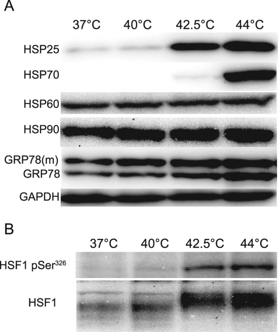
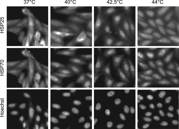
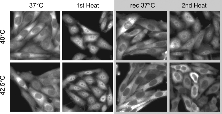
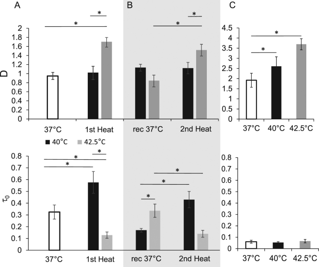
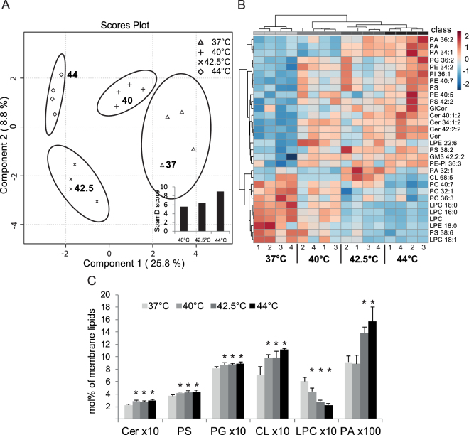
The current research on cellular heat stress management focuses on the roles of heat shock proteins (HSPs) and the proteostasis network under severe stress conditions. The mild, fever-type stress and the maintenance of membrane homeostasis are less well understood. Herein, we characterized the acute effect of mild, fever-range heat shock on membrane organization, and HSP synthesis and localization in two mammalian cell lines, to delineate the role of membranes in the sensing and adaptation to heat. A multidisciplinary approach combining ultrasensitive fluorescence microscopy and lipidomics revealed the molecular details of novel cellular "eustress", when cells adapt to mild heat by maintaining membrane homeostasis, activating lipid remodeling, and redistributing chaperone proteins. Notably, this leads to acquired thermotolerance in the complete absence of the induction of HSPs. At higher temperatures, additional defense mechanisms are activated, including elevated expression of molecular chaperones, contributing to an extended stress memory and acquired thermotolerance.


Conflict of interest statement

The authors declare that they have no competing interests.

Figures

Figure 1
Figure 1
The effect of temperature on HSP induction and HSF1 activation. (A) A GPI-mGFP–expressing CHO cell line was incubated for 20 min at the specified temperatures; the samples were prepared for western blotting after 6 h of recovery at 37 °C. (B) HSF1 phosphorylation was analyzed in samples after 20 min of heat treatment without recovery. See Supplementary Figure S5 and S6 for the full images of Fig. 1A,B, respectively.
Figure 2
Figure 2
The effect of heat treatment on the intracellular localization of HSPs in CHO cells. Representative images (n = 40) of CHO cells after a 20 min heat treatment at the indicated temperatures are shown.
Figure 3
Figure 3
ATT of CHO cells primed with different heat doses. A quantitative analysis of the results of colony formation assay is shown. CHO cells were primed for 20 min at the specified temperatures; after a 6 h recovery at 37 °C, the cells were exposed to 46 °C for 20 min. The mean values were normalized to controls which were kept at 37 °C throughout the duration of the experiments (n = 3; error bars represent SEM; *p < 0.05 indicates significance vs. time 0, Student’s t-test).
Figure 4
Figure 4
HSP25 distribution after two consecutive heat treatments of CHO cells. Representative images (n = 38) of HSP25 distribution in CHO cells at 37 °C, after a 20 min heat treatment at specified temperatures (1st heat); followed by 6 h recovery at 37 °C (rec 37 °C); and after a second, 20 min, heat treatment at specified temperatures (2nd heat).
Figure 5
Figure 5
The alteration of membrane diffusion of GPI-mGFP in live cells at different heat exposures. The diffusion coefficient (D) and confinement time (τ 0) of GPI-mGFP on the surface of CHO cells were measured by ImFCS (A) during a 15 min primary treatment (1st heat) at 37 °C, 40 °C, and 42.5 °C; (B) after a 6 h recovery at 37 °C (37 °C), and upon a 15 min second heating cycle (2nd heat) at the same temperature as in (A). (C) ImFCS analysis of PM sheets isolated from CHO cells, at 37 °C, 40 °C, or 42.5 °C. The values represent averages, and the error bars represent the standard error of mean (SEM) (n = 8, p < 0.05).
Figure 6
Figure 6
Characterization of lipidomes of GPI-mGFP–expressing CHO cells exposed to different levels of HS. (A) sPLS-DA and SoamD score (inset) of lipid molecular species. (B) Heatmap representation of hierarchical clustering of lipid profiles at 37 °C, 40 °C, 42.5 °C, and 44 °C. The top 30 most significant species were selected using ANOVA, Euclidean distance, and clustering algorithm Ward. The heat color-code represents normalized values (z-scores), ranging between [−2] (darkest shade of blue) and [+2] (darkest shade of red). Data from four independent experiments with control and HS-treated cells are shown. (C) Key features of HS-associated lipid remodeling of CHO lipidomes: ceramide (Cer), phosphatidylserine (PS), phosphatidylglycerol (PG), cardiolipin (CL), lysophosphatidylcholine (LPC), and phosphatidic acid (PA). The cells were either unstressed (37 °C) or stressed at 40 °C, 42.5 °C, or 44 °C, for 20 min. The values are expressed as mol% of membrane lipids (mean +SD), n = 4; *p < 0.05, (vs. 37 °C).

References

    1. Kültz D. Molecular and Evolutionary Basis of the Cellular Stress Response. Annu. Rev. Physiol. 2004;67:225–257. doi: 10.1146/annurev.physiol.67.040403.103635. - DOI - PubMed
    1. Selye, H. Stress without Distress. in Psychopathology of Human Adaptation 137–146 (Springer US, 1976). 10.1007/978-1-4684-2238-2_9
    1. Park HG, Han SI, Oh SY, Kang HS. Cellular responses to mild heat stress. Cell. Mol. Life Sci. 2005;62:10–23. doi: 10.1007/s00018-004-4208-7. - DOI - PMC - PubMed
    1. Velazquez JM, Lindquist S. hsp70: Nuclear concentration during environmental stress and cytoplasmic storage during recovery. Cell. 1984;36:655–662. doi: 10.1016/0092-8674(84)90345-3. - DOI - PubMed
    1. Horvath I, Multhoff G, Sonnleitner A, Vigh L. Membrane-associated stress proteins: More than simply chaperones. Biochim. Biophys. Acta - Biomembr. 2008;1778:1653–1664. doi: 10.1016/j.bbamem.2008.02.012. - DOI - PubMed

Substances