. 2017 Nov 15;7(1):15650.
doi: 10.1038/s41598-017-15834-3.

Sodium channel NaV1.3 is important for enterochromaffin cell excitability and serotonin release

Affiliations

Sodium channel NaV1.3 is important for enterochromaffin cell excitability and serotonin release

Peter R Strege et al. Sci Rep. .

Abstract

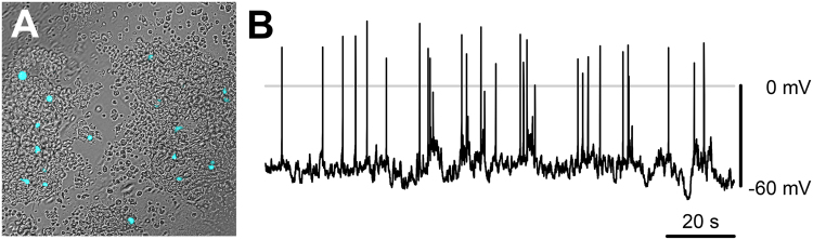
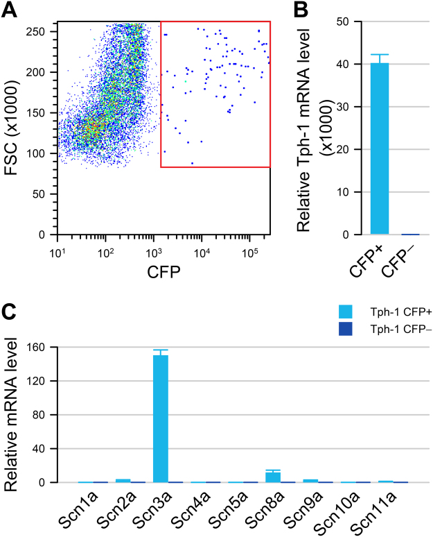
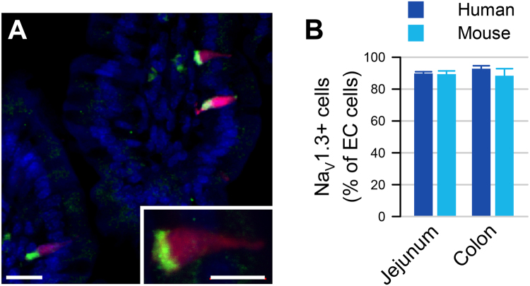
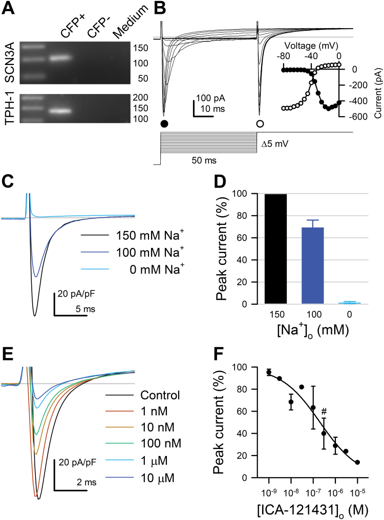
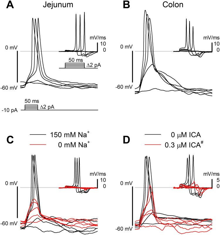
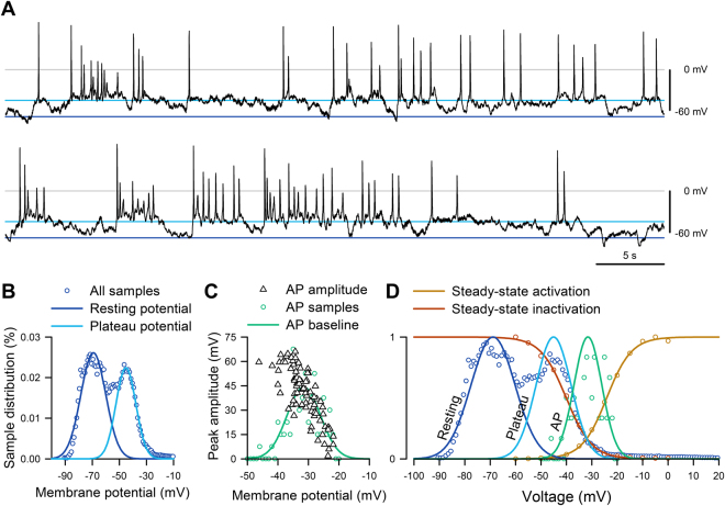
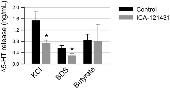
In the gastrointestinal (GI) epithelium, enterochromaffin (EC) cells are enteroendocrine cells responsible for producing >90% of the body's serotonin (5-hydroxytryptamine, 5-HT). However, the molecular mechanisms of EC cell function are poorly understood. Here, we found that EC cells in mouse primary cultures fired spontaneous bursts of action potentials. We examined the repertoire of voltage-gated sodium channels (NaV) in fluorescence-sorted mouse EC cells and found that Scn3a was highly expressed. Scn3a-encoded NaV1.3 was specifically and densely expressed at the basal side of both human and mouse EC cells. Using electrophysiology, we found that EC cells expressed robust NaV1.3 currents, as determined by their biophysical and pharmacologic properties. NaV1.3 was not only critical for generating action potentials in EC cells, but it was also important for regulating 5-HT release by these cells. Therefore, EC cells use Scn3a-encoded voltage-gated sodium channel NaV1.3 for electrical excitability and 5-HT release. NaV1.3-dependent electrical excitability and its contribution to 5-HT release is a novel mechanism of EC cell function.


Conflict of interest statement

The authors declare that they have no competing interests.

Figures

Figure 1
Figure 1
Primary EC cells have spontaneous electrical excitability. (A) Overlaid DIC/epifluorescence image of a primary colon culture from a Tph1-CFP mouse. CFP fluorescent cells (cyan) are EC cells. (B) Spontaneous electrical activity from a murine primary small bowel EC cell (whole cell current clamp).
Figure 2
Figure 2
Tph1-CFP EC cells highly express Scn3a. (A) Fluorescence-activated sorting of dissociated small bowel epithelial cells from Tph1-CFP mouse showing that 0.74% are CFP+ cells. (B) In the CFP+ fraction (cyan), Tph1 is highly expressed compared to CFP− (blue) cells. (C) Voltage-gated sodium channel α-subunit mRNA levels in CFP+ (cyan) and CFP− (blue) cells, showing a high expression of Scn3a in CFP+ EC cells but not in CFP− cells.
Figure 3
Figure 3
Scn3a-encoded NaV1.3 is specific to EC cells of mouse and human. (A) Expression of EC cell marker Tph-1 and voltage-gated sodium channel gene Scn3a by RNAseq in FACS-sorted Tph1-CFP EC cells from mouse small bowel. Red, TPH1-CFP; green, NaV1.3; blue, DAPI. Scale bar, 10 µm; inset, 5 µm. Inset, NaV1.3 is localized at the basal side of mouse EC cells. (B) Percentages of NaV1.3+/5HT+ cells out of all EC (5HT+) cells in human (blue) and NaV1.3+/CFP+ cells out of all EC cells (CFP+) in mouse (cyan) jejunum (left) and colon (right), as counted from immunohistochemistry data.
Figure 4
Figure 4
Primary cultured mouse small bowel EC cells express Scn3a and have fast voltage-gated inward currents that are selective for Na+ and inhibited by the NaV1.3 blocker ICA-121431. (A) Cropped single cell RT-PCR gel of Tph1-CFP+, Tph1-CFP- cells, or medium. (B) Representative traces of fast inward currents of voltage-clamped Tph1-CFP+ (EC) cells, elicited by a 2-step voltage ladder protocol (bottom). Inset, Peak currents of the traces shown in B, elicited during the activation (step 1, ●) or inactivation (step 2, ○) steps, plotted versus the voltage of step 1. (C) Representative traces of inward currents elicited by a step from −120 to −20 mV with 150 (black), 100 (blue), or 0 mM (cyan) extracellular [Na+]. Replacement of extracellular Na+ with N-methyl D-glucamine (NMDG+) diminishes (100 mM Na+) or eliminates (0 mM Na+) the fast inward currents. (D) Average peak Na+ current at the −20 mV test pulse with 100 mM [Na+]o (blue) or 0 mM [Na+]o (cyan), normalized to the peak Na+ current recorded with 150 mM [Na+]o (black) (n = 4). (E) Representative Na+ currents elicited from small bowel EC cells by steps from −120 to −20 mV with 0 (black) or 10−9 to 10−5 mol/L NaV1.3 blocker ICA-121431 (color gradient). (F) Dose-response of peak Na+ currents to ICA-121431 (IC50, 131 ± 54 nM; maximum block, 68 ± 7%, n = 4). Symbol (#) denotes 0.3 µM ICA-121431, the concentration used in Fig. 5D.
Figure 5
Figure 5
NaV1.3 is necessary for EC cell action potentials. (A,B) Typical action potentials from small bowel (A) and colon (B) EC cells, elicited by 50-ms depolarizing current steps (bottom). (C,D) Elicited EC cell action potentials were blocked by Na+ ion replacement (C) or NaV1.3 inhibition using ICA-121431 (300 nM, as highlighted by # in Fig. 4F). (D) Insets, first derivatives of the traces in each panel, showing the rate of change in membrane potential induced by the current injection protocol. The maximum depolarization rate is an approximate measure of the inward current through voltage-gated sodium channels.
Figure 6
Figure 6
EC cells fire spontaneous action potentials. (A) Current clamp recording of spontaneous electrical activity in the absence of current stimulus from a Tph1-CFP+ EC cell from mouse small bowel. In voltage clamp mode, this EC cell had 300 pA peak Na+ current, −24 mV half-maximal activation (V1/2A), and −41 mV half-maximal inactivation (V1/2I) (data not shown). (B) Sample distribution of the membrane potential (circles) from the full recording of panel A, fit with two Gaussian functions, showing separation into resting (blue) and plateau (cyan) potentials. (C) Peak amplitudes of action potentials (AP, triangles) versus the baseline prior to the firing of each AP. Distribution of AP (circles, n = 0–7 per 1-mV bin) was fit with a Gaussian function (green). (D) Action potential baseline distribution overlaps NaV1.3 window current. Normalized steady-state activation (red) or inactivation (yellow) currents from voltage-clamp (fit with Boltzmann functions), overlaying the normalized all-sample distribution of resting (blue) and plateau (cyan) potentials from (B) or AP baseline (green) from (C).
Figure 7
Figure 7
NaV1.3 contributes toward 5-HT release from EC cells. The change in release of 5-HT (Δ5-HT) from primary colonic EC cells was measured by ELISA following 20 min incubation with KCl (left),BDS-1 (middle) or butyrate (right), in the absence (black) or presence (gray) of NaV1.3 inhibitor ICA-121431.

References

    1. Cote F, et al. Disruption of the nonneuronal tph1 gene demonstrates the importance of peripheral serotonin in cardiac function. Proc. Natl. Acad. Sci. USA. 2003;100:13525–13530. doi: 10.1073/pnas.2233056100. - DOI - PMC - PubMed
    1. Mawe GM, Hoffman JM. Serotonin signalling in the gut–functions, dysfunctions and therapeutic targets. Nature Reviews: Gastroenterology and Hepatology. 2013;10:473–486. - PMC - PubMed
    1. Heredia DJ, et al. Important role of mucosal serotonin in colonic propulsion and peristaltic reflexes: in vitro analyses in mice lacking tryptophan hydroxylase 1. J. Physiol. 2013;591:5939–5957. doi: 10.1113/jphysiol.2013.256230. - DOI - PMC - PubMed
    1. Heredia DJ, Dickson EJ, Bayguinov PO, Hennig GW, Smith TK. Localized release of serotonin (5-hydroxytryptamine) by a fecal pellet regulates migrating motor complexes in murine colon. Gastroenterology. 2009;136:1328–1338. doi: 10.1053/j.gastro.2008.12.010. - DOI - PMC - PubMed
    1. Keating DJ, Spencer NJ. Release of 5-hydroxytryptamine from the mucosa is not required for the generation or propagation of colonic migrating motor complexes. Gastroenterology. 2010;138(659–670):670 e651–652. - PubMed

MeSH terms