

doi: 10.4103/ijnm.IJNM_93_17.
Utility of 18F-Fluorodeoxyglucose Positron Emission Tomography/Magnetic Resonance Imaging in the Diagnosis of Cardiac Paraganglioma
Affiliations
- PMID: 29142369
- PMCID: PMC5672773
- DOI: 10.4103/ijnm.IJNM_93_17
Item in Clipboard
Utility of 18F-Fluorodeoxyglucose Positron Emission Tomography/Magnetic Resonance Imaging in the Diagnosis of Cardiac Paraganglioma
Indian J Nucl Med.
2017 Oct-Dec.
Abstract
Cardiac paragangliomas are rare tumors of neural crest origin, most frequently seen in the left atrium. There are mixed opinions regarding the most appropriate imaging study for diagnosis and evaluation. We describe the novel utility of 18-F-Fluorodeoxyglucose positron emission tomography/magnetic resonance imaging in the case of a 42-year-old male with cardiac paraganglioma.
Keywords: 18F-Fluorodeoxyglucose positron emission tomography/magnetic resonance imaging; cardiac paraganglioma; neuroendocrine tumor.
Conflict of interest statement
There are no conflicts of interest.
Figures
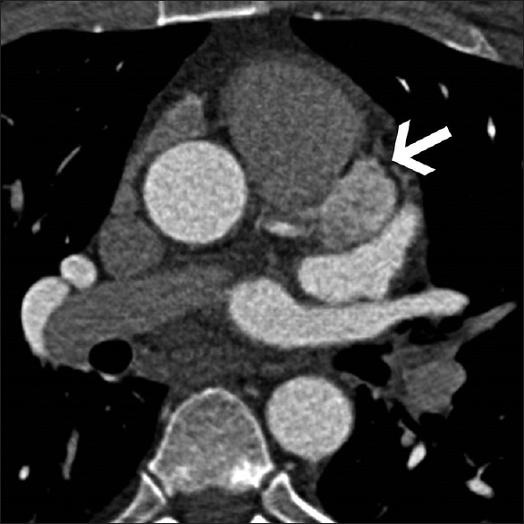
Axial intravenous contrast-enhanced computed tomography image shows an intensely enhancing well-defined mass (arrow) between the left superior pulmonary vein and the pulmonary artery and abutting the left anterior descending artery
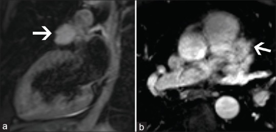
Hybrid 18F-Fluorodeoxyglucose positron emission tomography/magnetic resonance (Philips Ingenuity TF PET/MR Philips Healthcare, Andover, MA, USA) was performed. (a) Vertical long axis T2-weighted magnetic resonance image shows a hyperintense epicardial mass measuring 3 cm × 1.8 cm abutting the left anterior descending artery. (b) Axial postcontrast T1 fat suppressed magnetic resonance imaging shows intense contrast enhancement of the mass
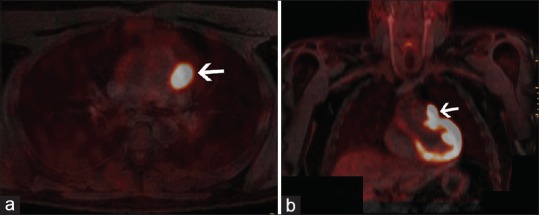
Axial (a) and coronal (b) hybrid 18F-Fluorodeoxyglucose positron emission tomography/magnetic resonance images of the same patient show the mass to be intensely hypermetabolic with standard uptake value of 16. No other hypermetabolic mass is seen to suggest metastatic disease
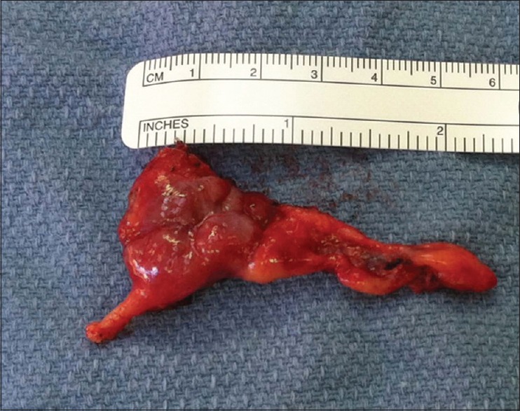
Gross surgical specimen of faint blue and tan-red muscular soft tissue with attached tan-yellow soft fatty tissue measuring 2.9 cm × 2.8 cm × 1.9 cm
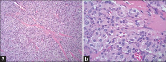
Histopathological image at 100× magnification (a) a tumor with classic Zellballen cell nests with rich vascular network and intervening fibrous bands. The same histopathological image at 400× (b) moderate nuclear atypia with “salt-and-pepper” chromatin pattern. This was consistent with paraganglioma
References
-
- Butany J, Nair V, Naseemuddin A, Nair GM, Catton C, Yau T. Cardiac tumours: Diagnosis and management. Lancet Oncol. 2005;6:219–28. - PubMed
-
- Orringer MB, Sisson JC, Glazer G, Shapiro B, Francis I, Behrendt DM, et al. Surgical treatment of cardiac pheochromocytomas. J Thorac Cardiovasc Surg. 1985;89:753–7. - PubMed
-
- Yadav PK, Baquero GA, Malysz J, Kelleman J, Gilchrist IC. Cardiac paraganglioma. Circ Cardiovasc Interv. 2014;7:851–6. - PubMed
-
- Wang JG, Han J, Jiang T, Li YJ. Cardiac paragangliomas. J Card Surg. 2015;30:55–60. - PubMed
LinkOut - more resources
Full Text Sources
Other Literature Sources
