

Higher PD-1 expression concurrent with exhausted CD8+ T cells in patients with de novo acute myeloid leukemia
- PMID: 29142466
- PMCID: PMC5677131
- DOI: 10.21147/j.issn.1000-9604.2017.05.11
Higher PD-1 expression concurrent with exhausted CD8+ T cells in patients with de novo acute myeloid leukemia
Abstract
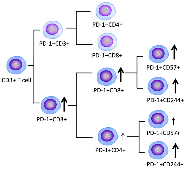
Objective: To investigate the association between the T cell inhibitory receptor programmed death 1 (PD-1) and T cell exhaustion status in T cells from patients with de novo acute myeloid leukemia (AML) and AML in complete remission (CR).
Methods: Surface expression of PD-1 and the exhaustion and immunosenescence markers CD244 and CD57 on CD3+, CD4+ and CD8+ T cells from peripheral blood samples from 20 newly diagnosed, untreated AML patients and 10 cases with AML in CR was analyzed by flow cytometry. Twenty-three healthy individuals served as control.
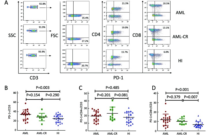
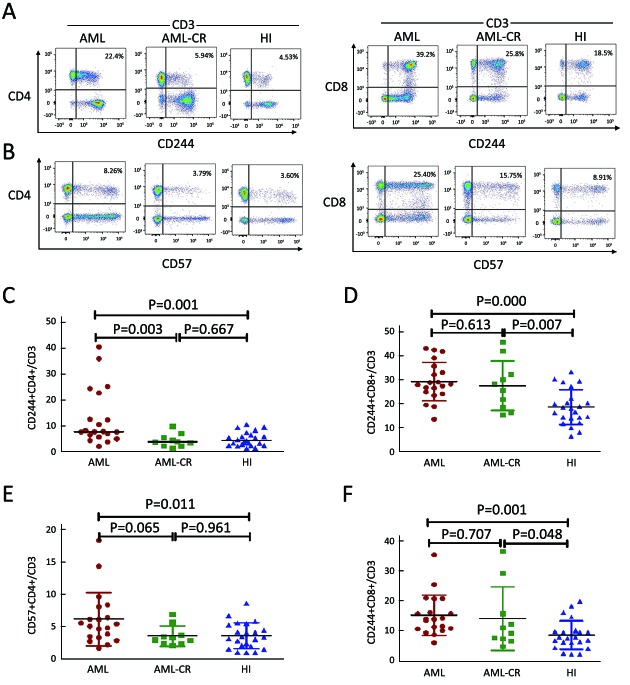
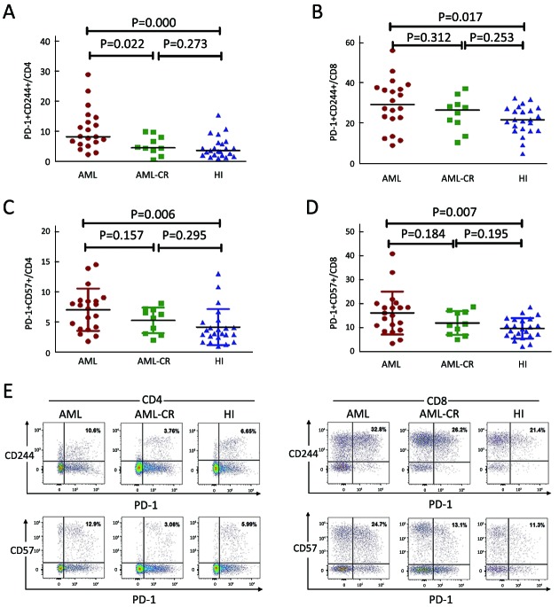
Results: A significantly higher percentage of PD-1+ cells were found for CD3+ T cells in the de novo AML group compared with healthy controls. In addition, an increased level of PD-1+CD8+ T cells, but not PD-1+CD4+, was found for CD3+ T cells in the de novo AML and AML-CR samples. A higher percentage of CD244+CD4+, CD244+CD8+, CD57+CD4+ and CD57+CD8+ T cells was found in CD3+ T cells in samples from those with de novo AML compared with those from healthy controls. Strong increased PD-1+CD244+ and PD-1+CD57+ co-expression was found for CD4+ and CD8+ T cells in the de novo AML group compared with healthy controls.
Conclusions: We characterized the major T cell defects, including co-expression of PD-1 and CD244, CD57-exhausted T cells in patients with de novo AML, and found a particular influence on CD8+ T cells, suggesting a poor anti-leukemia immune response in these patients.
Keywords: Acute myeloid leukemia; PD-1; T cell exhaustion.
Figures
Similar articles
-
Increased PD-1+Tim-3+ exhausted T cells in bone marrow may influence the clinical outcome of patients with AML.Biomark Res. 2020 Feb 13;8:6. doi: 10.1186/s40364-020-0185-8. eCollection 2020. Biomark Res. 2020. PMID: 32082573 Free PMC article.
-
Increased TOX expression concurrent with PD-1, Tim-3, and CD244 expression in T cells from patients with acute myeloid leukemia.Cytometry B Clin Cytom. 2022 Mar;102(2):143-152. doi: 10.1002/cyto.b.22049. Epub 2021 Dec 16. Cytometry B Clin Cytom. 2022. PMID: 34913594
-
Increasing Tim-3+CD244+, Tim-3+CD57+, and Tim-3+PD-1+ T cells in patients with acute myeloid leukemia.Asia Pac J Clin Oncol. 2020 Jun;16(3):137-141. doi: 10.1111/ajco.13304. Epub 2020 Feb 7. Asia Pac J Clin Oncol. 2020. PMID: 32030888
-
[Biological properties and sensitivity to induction therapy of differentiated cells expressing atypical immunophenotype in acute leukemia of children].Folia Med Cracov. 2001;42(3):5-80. Folia Med Cracov. 2001. PMID: 12353422 Review. Polish.
-
Immunoediting in acute myeloid leukemia: Reappraising T cell exhaustion and the aberrant antigen processing machinery in leukemogenesis.Heliyon. 2024 Oct 25;10(21):e39731. doi: 10.1016/j.heliyon.2024.e39731. eCollection 2024 Nov 15. Heliyon. 2024. PMID: 39568858 Free PMC article. Review.
Cited by
-
Clinical significance of T helper cell subsets in the peripheral blood and bone marrow of patients with multiple myeloma.Front Immunol. 2024 Sep 11;15:1445530. doi: 10.3389/fimmu.2024.1445530. eCollection 2024. Front Immunol. 2024. PMID: 39324138 Free PMC article.
-
T cell receptor-engineered T cells for leukemia immunotherapy.Cancer Cell Int. 2019 Jan 3;19:2. doi: 10.1186/s12935-018-0720-y. eCollection 2019. Cancer Cell Int. 2019. PMID: 30622438 Free PMC article. Review.
-
Increased PD-1+Tim-3+ exhausted T cells in bone marrow may influence the clinical outcome of patients with AML.Biomark Res. 2020 Feb 13;8:6. doi: 10.1186/s40364-020-0185-8. eCollection 2020. Biomark Res. 2020. PMID: 32082573 Free PMC article.
-
Extensive exploration of T cell heterogeneity in cancers by single cell sequencing.Chin J Cancer Res. 2019 Apr;31(2):410-418. doi: 10.21147/j.issn.1000-9604.2019.02.15. Chin J Cancer Res. 2019. PMID: 31156311 Free PMC article.
-
TIM-3 teams up with PD-1 in cancer immunotherapy: mechanisms and perspectives.Mol Biomed. 2025 May 7;6(1):27. doi: 10.1186/s43556-025-00267-6. Mol Biomed. 2025. PMID: 40332725 Free PMC article. Review.
References
LinkOut - more resources
Full Text Sources
Other Literature Sources
Research Materials