. 2017 Oct 28;23(40):7253-7264.
doi: 10.3748/wjg.v23.i40.7253.

Prostaglandin E1 protects hepatocytes against endoplasmic reticulum stress-induced apoptosis via protein kinase A-dependent induction of glucose-regulated protein 78 expression

Affiliations

Prostaglandin E1 protects hepatocytes against endoplasmic reticulum stress-induced apoptosis via protein kinase A-dependent induction of glucose-regulated protein 78 expression

Fang-Wan Yang et al. World J Gastroenterol. .

Abstract

Aim: To investigate the protective effect of prostaglandin E1 (PGE1) against endoplasmic reticulum (ER) stress-induced hepatocyte apoptosis, and to explore its underlying mechanisms.

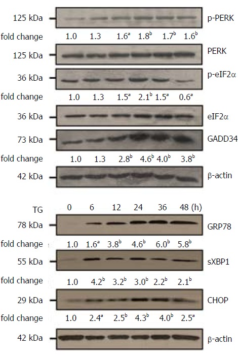
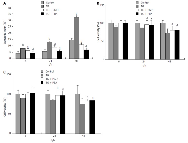
Methods: Thapsigargin (TG) was used to induce ER stress in the human hepatic cell line L02 and hepatocarcinoma-derived cell line HepG2. To evaluate the effects of PGE1 on TG-induced apoptosis, PGE1 was used an hour prior to TG treatment. Activation of unfolded protein response signaling pathways were detected by western blotting and quantitative real-time RT-PCR. Apoptotic index and cell viability of L02 cells and HepG2 cells were determined with flow cytometry and MTS [3-(4,5-dimethylthiazol-2-yl)-5-(3-carboxymethoxyphenyl)-2-(4-sulfophenyl)-2H-tetrazolium] assay.

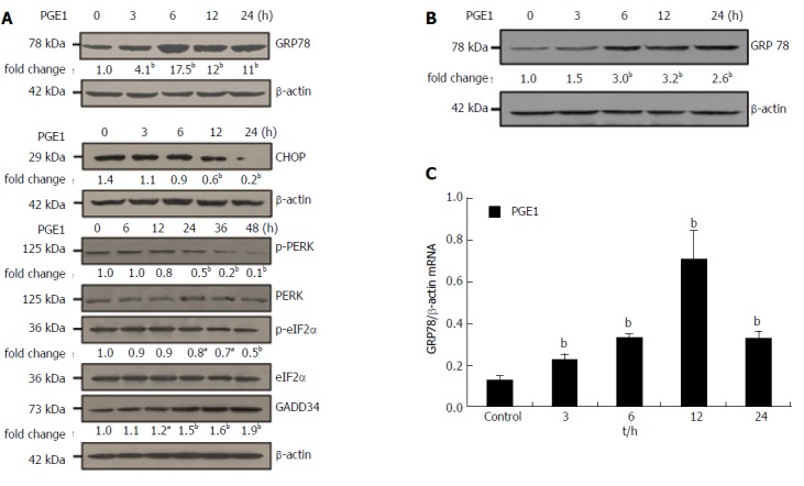
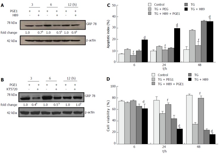
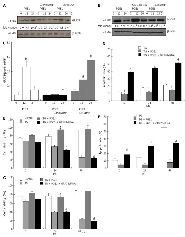
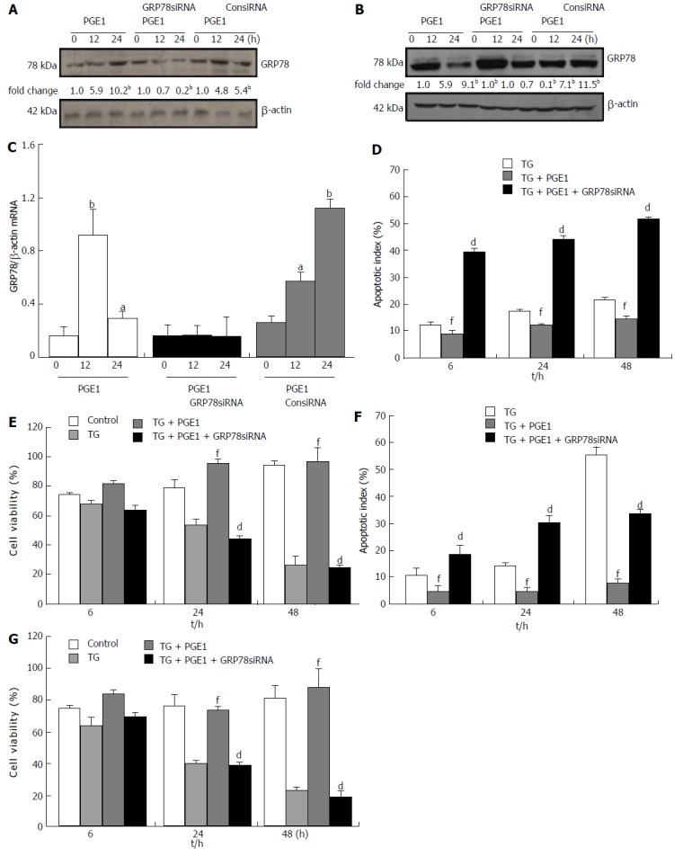
Results: Pretreatment with 1 μmol/L PGE1 protected against TG-induced apoptosis in both L02 cells and HepG2 cells. PGE1 enhanced the TG-induced expression of C/EBP homologous protein (CHOP), glucose-regulated protein (GRP) 78 and spliced X box-binding protein 1 at 6 h. However, it attenuated their expressions after 24 h. PGE1 alone induced protein and mRNA expressions of GRP78; PGE1 also induced protein expression of DNA damage-inducible gene 34 and inhibited the expressions of phospho-PKR-like ER kinase, phospho-eukaryotic initiation factor 2α and CHOP. Treatment with protein kinase A (PKA)-inhibitor H89 or KT5720 blocked PGE1-induced up-regulation of GRP78. Further, the cytoprotective effect of PGE1 on hepatocytes was not observed after blockade of GRP78 expression by H89 or small interfering RNA specifically targeted against human GRP78.

Conclusion: Our study demonstrates that PGE1 protects against ER stress-induced hepatocyte apoptosis via PKA pathway-dependent induction of GRP78 expression.

Keywords: Apoptosis; Endoplasmic reticulum stress; Glucose-regulated protein 78; Hepatocytes; Protein kinase A; Thapsigargin.


Conflict of interest statement

Conflict-of-interest statement: The authors declare that they have no conflicts interest related to this study.

Figures

Figure 1
Figure 1
TG induced endoplasmic reticulum stress in L02 cells. L02 cells were treated with 1 μmol/L TG for 6, 12, 24, 36 and 48 h. Expressions of p-PERK, p-eIF2α, GADD34, GRP78, sXBP1 and CHOP were assessed by western blotting. Representative blots from three independent experiments are presented. The results of densitometric analysis are presented as a fold-change compared to that at 0 h (aP < 0.05; bP < 0.01). CHOP: C/EBP homologous protein; ER: Endoplasmic reticulum; GADD34: Growth arrest and DNA damage-inducible gene34; GRP78: Glucose-regulated protein 78; p-eIF2α: Phospho-eukaryotic translation initiation factor-2α; p-PERK: Phospho-PKR-like ER kinase; sXBP1: Spliced X box-binding protein1; TG: Thapsigargin.
Figure 2
Figure 2
Prostaglandin E1 protected against thapsigargin-induced apoptosis in both L02 cells and HepG2 cells. L02 cells and HepG2 cells were pretreated with PGE1 or PBA for 1 h and treated with a final concentration of 1 μmol/L TG for 6, 24 and 48 h. A: Apoptotic index of L02 cells was determined by flow cytometry. Histograms represent mean ± SD of five separate experiments, each of which was performed in triplicate. bP < 0.01 vs control at the same time point; dP < 0.01 vs TG at the same time point; B and C: Cell viability of L02 cells and HepG2 cells was determined by MTS assay. The absorbance was measured at 490 nm and cell viability was normalized as a percentage of control. Histograms represent mean ± SD of five separate experiments, each of which was performed in triplicate. dP < 0.01 vs TG at the same time point. MTS: [3-(4,5-dimethylthiazol-2-yl)-5-(3-carboxymethoxyphenyl)-2-(4-sulfophenyl)-2H-tetrazolium]; PBA: 4-phenylbutyric acid; PGE1: Prostaglandin E1; TG: Thapsigargin.
Figure 3
Figure 3
Prostaglandin E1 promoted rapid recovery of endoplasmic reticulum stress. L02 cells were pretreated with 1 μmol/L PGE1 for 1 h and at a final concentration of 1 μmol/L TG for 6, 24 and 48 h. Expressions of GRP78, CHOP and sXBP1) were assessed on western blotting. One representative blot each of three individual experiments is presented. The results of densitometric analysis are presented as a fold-change compared to those of TG at the same time point (aP < 0.05, bP < 0.01). CHOP: C/EBP homologous protein; ER: Endoplasmic reticulum; GRP78: Glucose-regulated protein 78; PGE1: Prostaglandin E1; sXBP1: Spliced X box-binding protein 1; TG: Thapsigargin.
Figure 4
Figure 4
Prostaglandin E1 induced glucose-regulated protein 78 protein and mRNA expressions. To determine GRP78 and CHOP protein or mRNA expressions, L02 cells and HepG2 cells were treated with 1 μmol/L PGE1 for 3, 6, 12 and 24 h; for detecting GADD34 and the p-PERK and p-eIF2α, L02 cells were treated with 1 μmol/L PGE1 for 6, 12, 24, 36 and 48 h. A: Expressions of GRP78, CHOP, GADD34, p-PERK and p-eIF2α in L02 cells were assessed by western blotting. One representative blot each from the three individual experiments is presented. The results of densitometric analysis are presented as a fold-change compared to those at 0 h (aP < 0.05, bP < 0.01); B: Expression of GRP78 in HepG2 cells was assessed by western blotting. One representative blot each from the three individual experiments is presented. The results of densitometric analysis are presented as a fold-change compared to those at 0 h (bP < 0.01); C: mRNA expression of GRP78 in L02 cells was assessed by quantitative real-time PCR (E). Histograms represent mean ± SD of three experiments (bP < 0.01 vs those at 0 h). CHOP: C/EBP homologous protein; ER: Endoplasmic reticulum; GADD34: Growth arrest and DNA damage-inducible gene 34; GRP78: Glucose-regulated protein 78; p-eIF2α: Phospho-eukaryotic translation initiation factor-2α; PGE1: Prostaglandin E1; p-PERK: Phospho-PKR-like ER kinase; TG: Thapsigargin.
Figure 5
Figure 5
Prostaglandin E1 induced glucose-regulated protein 78 expression and protected L02 cells against endoplasmic reticulum stress-induced apoptosis via a protein kinase a-dependent pathway. A and B: L02 cells were pretreated with or without PKA inhibitor H89 (10 μmol/L) or KT5720 (1 μmol/L), and then 1 μmol/L PGE1 for 3, 6 and 12 h. The expressions of GRP78 were detected by western blotting. One representative blot each from three independent experiments is presented. The results of densitometric analysis are presented as a fold-change compared to those of PGE1 at the same time points (bP < 0.01); C: L02 cells were pretreated with or without 10 μmol/L H89, and then 1 μmol/L PGE1 and a final concentration of 1 μmol/L TG for 6, 24 and 48 h. The apoptotic index was determined by flow cytometry. Histograms represent mean ± SD of five separate experiments, each of which was performed in triplicate. (dP < 0.01 vs those of TG + PGE1 at the same time point; fP < 0.01 vs those of TG at the same time point); D: L02 cells were pretreated with or without 10 μmol/L H89, and then 1 μmol/L PGE1 and a final concentration of 1 μmol/L TG for 6, 24 and 48 h. Cell viability of L02 cells was determined by MTS assay. Histograms represent mean ± SD of five separate experiments, each of which was performed in triplicate (dP < 0.01 vs those of TG + PGE1 at the same time point; fP < 0.01 vs those of TG at the same time point). GRP78: Glucose-regulated protein 78; MTS: [3-(4,5-dimethylthiazol-2-yl)-5-(3-carboxymethoxyphenyl)-2-(4-sulfophenyl)-2H-tetrazolium]; PGE1: Prostaglandin E1; PKA: Protein kinase A; TG: Thapsigargin.
Figure 6
Figure 6
Prostaglandin E1 protected against endoplasmic reticulum stress-induced apoptosis via induction of glucose-regulated protein 78 expression in both L02 and HepG2 cells. L02 and HepG2 cells were transfected with either siRNA scramble control (ConsiRNA) or siRNA against human GRP78 (GRP78 siRNA) for 48 h. The cells were treated with 1 μmol/L PGE1 for 12 h and 24 h. A and B: Expressions of GRP78 in L02 cells and in HepG2 cells were detected by western blotting. One representative blot each from the three individual experiments is presented. The results of densitometric analysis are presented as a fold-change compared to those at 0 h (bP < 0.01); C: mRNA expression of GRP78 in L02 cells was detected by real-time PCR. Histograms represent mean ± SD of three experiments (aP < 0.05; bP < 0.01 vs those at 0 h). D and F: Apoptotic indices of L02 cells and HepG2 cells were determined by flow cytometry. Histograms represent mean ± SD of five independent experiments, each of which was performed in triplicate (dP < 0.01 vs those of TG + PGE1 at the same time point; fP < 0.01 vs those of TG at the same time point); E and G: Cell viability of L02 cells and HepG2 cells was determined by MTS assay. Histograms represent mean ± SD of five independent experiments, each of which was performed in triplicate (dP < 0.01 vs those of TG + PGE1 and fP < 0.01 vs those of TG at the same time point). GRP78: Glucose-regulated protein 78; PGE1: Prostaglandin E1; MTS: [3-(4,5-dimethylthiazol-2-yl)-5-(3-carboxymethoxyphenyl)-2-(4-sulfophenyl)-2H-tetrazolium]; siRNA: Small interfering RNA; TG: Thapsigargin.

Similar articles

Cited by

References

    1. Guicciardi ME, Malhi H, Mott JL, Gores GJ. Apoptosis and necrosis in the liver. Compr Physiol. 2013;3:977–1010. - PMC - PubMed
    1. Malhi H, Gores GJ. Cellular and molecular mechanisms of liver injury. Gastroenterology. 2008;134:1641–1654. - PMC - PubMed
    1. Brenner C, Galluzzi L, Kepp O, Kroemer G. Decoding cell death signals in liver inflammation. J Hepatol. 2013;59:583–594. - PubMed
    1. Sano R, Reed JC. ER stress-induced cell death mechanisms. Biochim Biophys Acta. 2013;1833:3460–3470. - PMC - PubMed
    1. Zha BS, Zhou H. ER Stress and Lipid Metabolism in Adipocytes. Biochem Res Int. 2012;2012:312943. - PMC - PubMed

MeSH terms