. 2015 May 1;6(5):2782-false.
doi: 10.1039/c4sc03739a. Epub 2015 Feb 9.

In vitro and in vivo comparative and competitive activity-based protein profiling of GH29 α-l-fucosidases

Affiliations

In vitro and in vivo comparative and competitive activity-based protein profiling of GH29 α-l-fucosidases

Jianbing Jiang et al. Chem Sci. .

Abstract

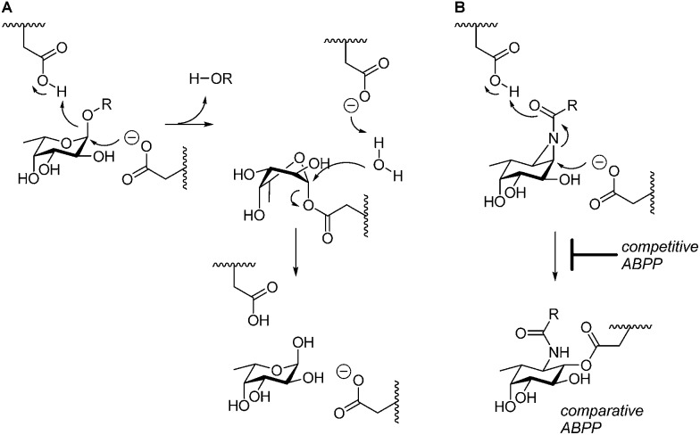
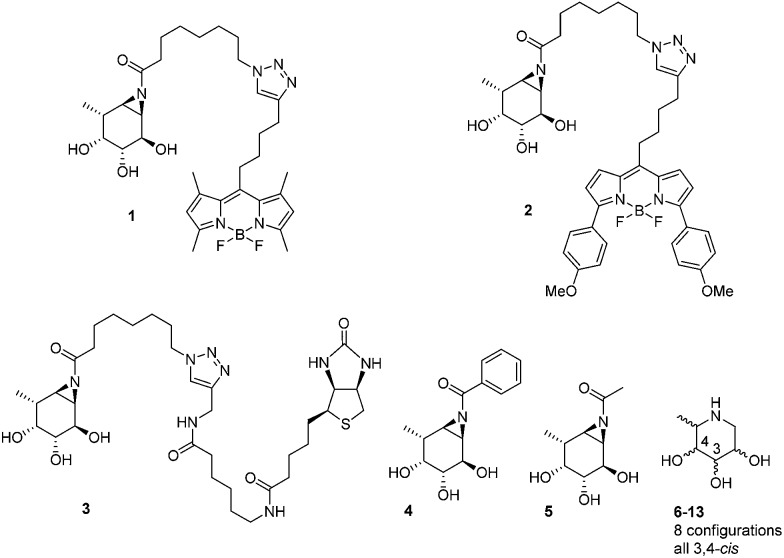
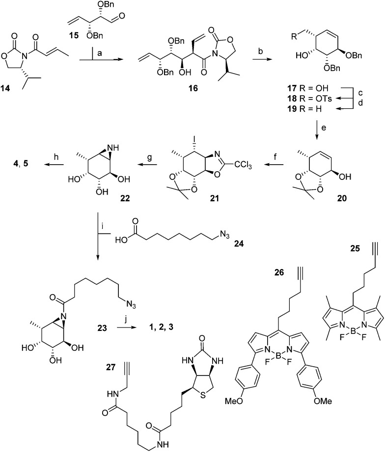
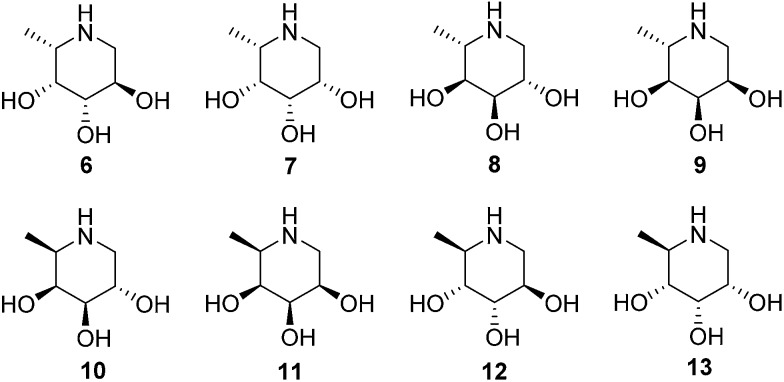
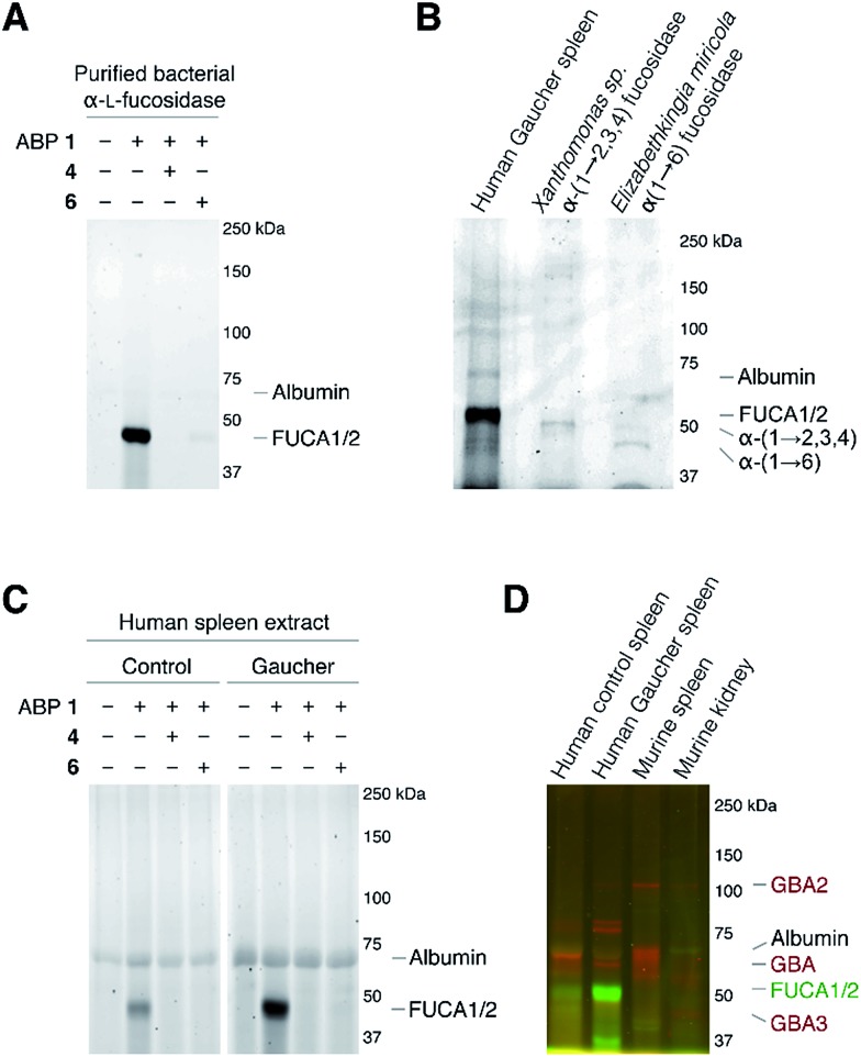
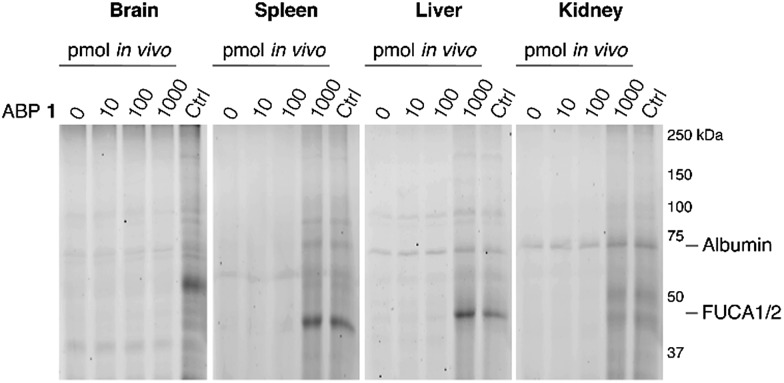
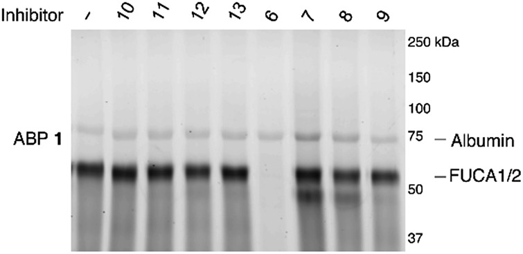
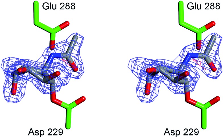
GH29 α-l-fucosidases catalyze the hydrolysis of α-l-fucosidic linkages. Deficiency in human lysosomal α-l-fucosidase (FUCA1) leads to the recessively inherited disorder, fucosidosis. Herein we describe the development of fucopyranose-configured cyclophellitol aziridines as activity-based probes (ABPs) for selective in vitro and in vivo labeling of GH29 α-l-fucosidases from bacteria, mice and man. Crystallographic analysis on bacterial α-l-fucosidase confirms that the ABPs act by covalent modification of the active site nucleophile. Competitive activity-based protein profiling identified l-fuconojirimycin as the single GH29 α-l-fucosidase inhibitor from eight configurational isomers.


Figures

Fig. 1
Fig. 1. (A) Double-displacement mechanism of retaining α-l-fucosidases. (B) Comparative and competitive activity-based profiling of GH29 α-l-fucosidases presented here.
Fig. 2
Fig. 2. Structures of inhibitors and ABPs presented in this study.
Scheme 1
Scheme 1. Synthesis of aziridine ABPs 1, 2, 3 and inhibitors 4, 5. Reagents and conditions: (a) DBBT, Et3N, CH2Cl2, –78 °C, 71%; (b) (i) LiBH4, THF, 83%; (ii) Grubbs 2nd generation, CH2Cl2, 95%; (c) p-TsCl, Et3N, CH2Cl2, 87%; (d) LiAlH4, THF, 0 °C to rt, 87%; (e) (i) Li, NH3 (l), THF, –60 °C, 73%, (ii) 2,2-dimethoxypropane, CSA, rt, 60%; (f) (i) CCl3CN, DBU, CH2Cl2, rt; (ii) NaHCO3, I2, H2O, rt, 46%; (g) (i) 37% HCl (aq.), MeOH, 60 °C; (ii) NaHCO3, MeOH, rt, 65%; (h) EEDQ, benzoic acid or acetic acid, DMF, 0 °C, 7% 4, 28% 5; (i) EEDQ, 24, DMF, 0 °C 25%; (j) CuSO4, sodium ascorbate, 25, 26 or 27, rt, 19% 1, 12% 2, 13% 3.
Fig. 3
Fig. 3. Structures of 1-deoxy-l-fuconojirimycin 6 and configurational isomers 7–13.
Scheme 2
Scheme 2. Synthesis of 1-deoxy-l-fuconojirimycin 6. Reagents and conditions: (a) (i) Et2O, DIBAL-H, –80 °C, (ii) MeOH, –90 °C, (iii) amine 29, NaOMe, (iv) NaBH4, –15 °C to rt, 88%; (b) Boc2O, 50 °C, 100%; (c) Grubbs 1st generation, CH2Cl2, 88%; (d) (i) K2OSO4·2H2O, NMO, acetone–H2O (1 : 1), –10 °C, (ii) Ac2O, pyridine, DMAP, 0 °C, 88% (33 : 34 = 1 : 3); (e) (i) K2CO3, MeOH, (ii) TBAF, THF, (iii) 6M HCl, MeOH, 71%.
Fig. 4
Fig. 4. In vitro activity-based protein profiling of GH29 α-l-fucosidases. (A) Labeling with ABP 1 of recombinant α-l-fucosidase from Bacteroides thetaiotaomicron 2970. (B) In vitro labeling of lysate of spleen from a Gaucher disease patient, α-(1-2,3,4)-fucosidase from Xanthomonas sp. and α-(1-6)-fucosidase from Elizabethkingia miricola with ABP 2. (C) In vitro labeling of human healthy and Gaucher disease spleen. (D) Direct labeling of GH29 α-l-fucosidases with green-fluorescent ABP 1 and retaining β-glucosidases GBA, GBA2 and GBA3 with red-fluorescent JJB75. The location of albumin autofluorescence is designated on each gel.
Fig. 5
Fig. 5. In vivo labeling of α-l-fucosidases in mice with various concentrations of ABP 1 during 2 hours. In vivo labeling compared to maximal labeling with excess ABP 1 of matched homogenates (Ctrl). The location of albumin autofluorescence is designated on each gel.
Fig. 6
Fig. 6. Competitive ABPP on recombinant FUCA1 with deoxyfuconojirimycin 6 and configurational analogues 7–13 towards α-l-fucosidases, with ABP 1 labeling as readout.
Fig. 7
Fig. 7. Crystal structure of α-l-fucosidase from Bacteroides thetaiotaomicron in complex with 5. Catalytic residues are annotated: Asp 229 (nucleophile) and Glu 288 (acid/base). Electron density displayed is FoFc density from phases calculated prior to the inclusion of 5 in refinement, contoured at 3σ. Figure was prepared using CCP4MG.

References

    1. Intra J., Perotti M.-E., Pavesi G., Horner D. Gene. 2007;392:34–46. - PubMed
    1. Lombard V., Golaconda Ramulu H., Drula E., Coutinho P. M., Henrissat B. Nucleic Acids Res. 2014;42:D490–D495. - PMC - PubMed
    1. Liu S. W., Chen C. S., Chang S. S., Mong K. K. T., Lin C. H., Chang C. W., Tang C. Y., Li Y. K. Biochemistry. 2009;48:110–120. - PubMed
    1. Guillotin L., Lafite P., Daniellou R. Biochemistry. 2014;53:1447–1455. - PubMed
    1. Cobucci-Ponzano B., Mazzone M., Rossi M., Moracci M. Biochemistry. 2005;44:6331–6342. - PubMed

LinkOut - more resources