. 2015 Aug 1;6(8):4756-4766.
doi: 10.1039/c5sc00560d. Epub 2015 May 22.

Integrating proteomics with electrochemistry for identifying kinase biomarkers

Affiliations

Integrating proteomics with electrochemistry for identifying kinase biomarkers

Einav Amit et al. Chem Sci. .

Abstract

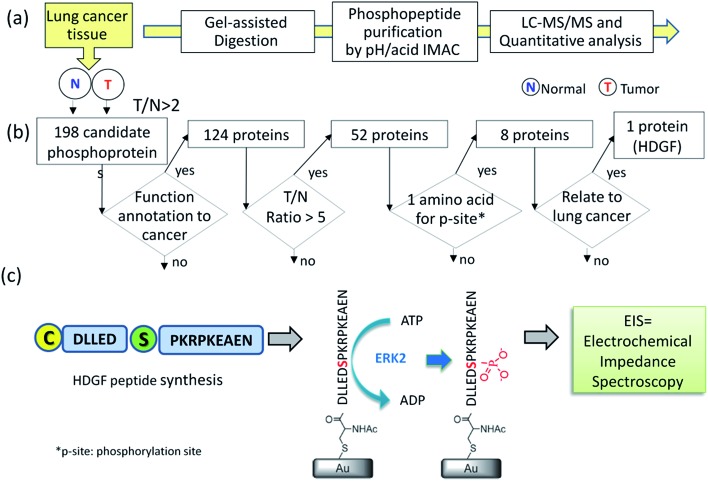
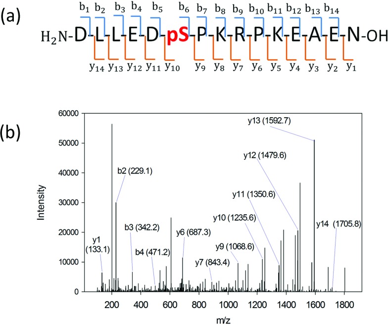
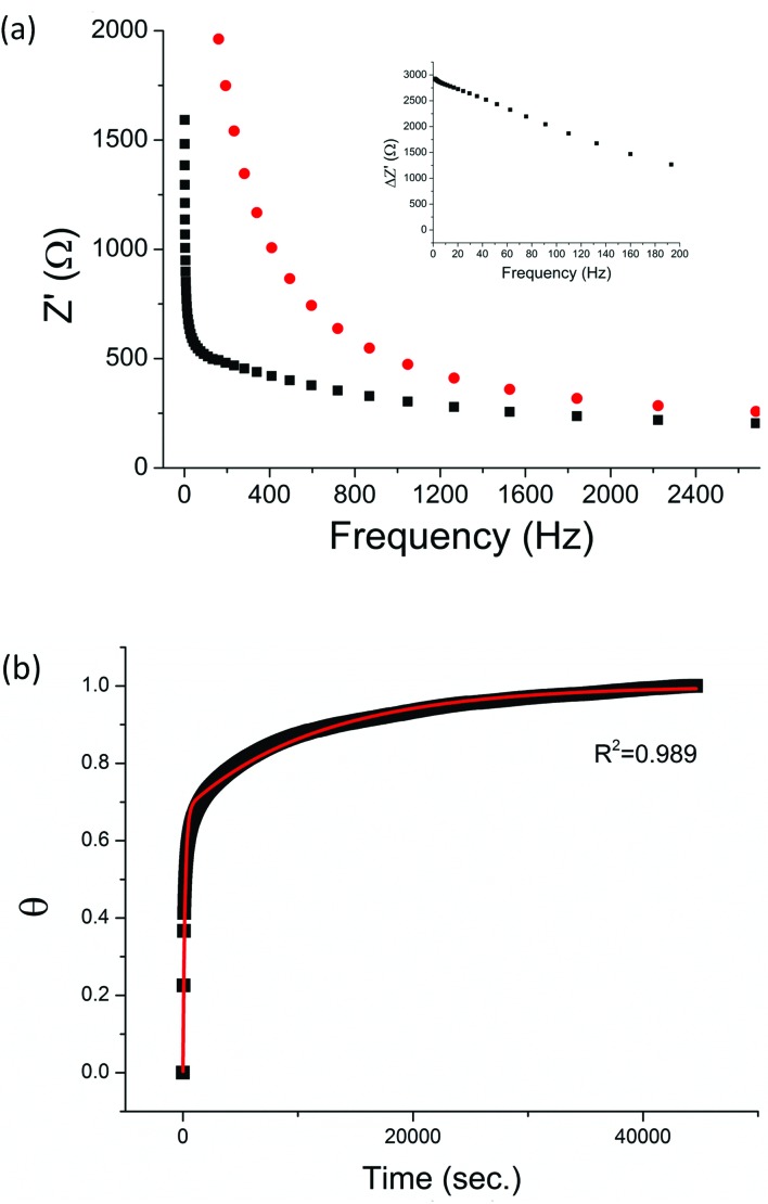
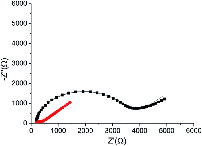
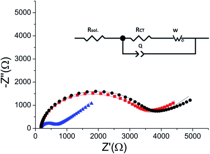
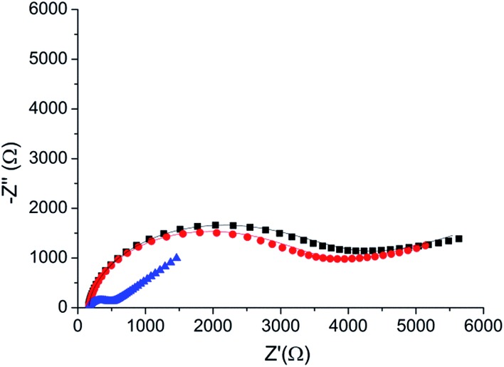
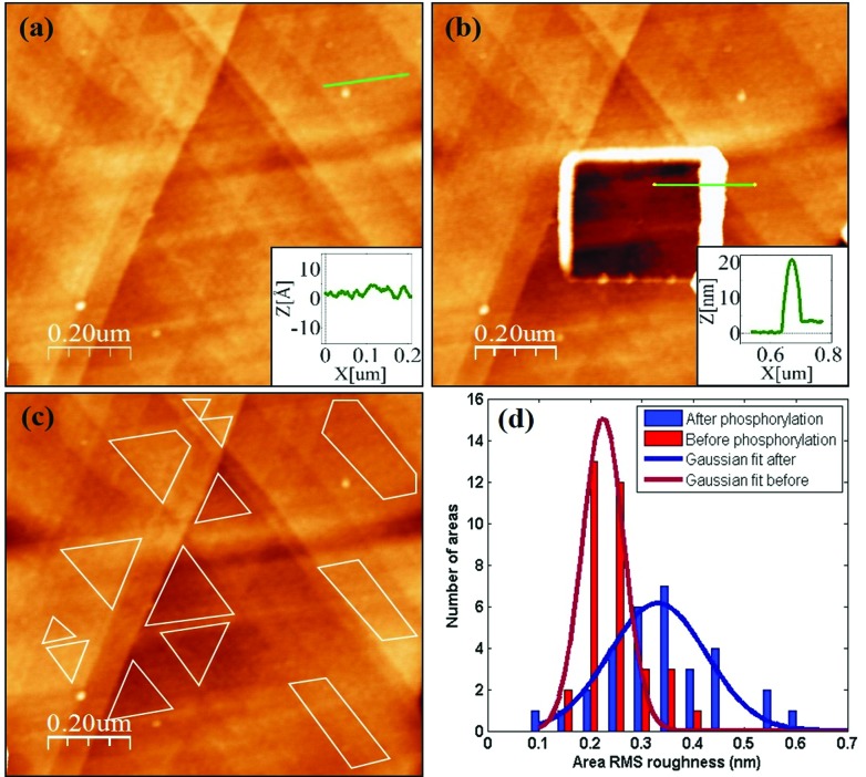
We present an integrated approach for highly sensitive identification and validation of substrate-specific kinases as cancer biomarkers. Our approach combines phosphoproteomics for high throughput cancer-related biomarker discovery from patient tissues and an impedimetric kinase activity biosensor for sensitive validation. Using non-small-cell lung cancer (NSCLC) as a proof-of-concept study, label-free quantitative phosphoproteomic analysis of a pair of cancerous and its adjacent normal tissues revealed 198 phosphoproteins that are over-phosphorylated in NSCLC. Among the differentially regulated phosphorylation sites, the most significant alteration was in residue S165 in the Hepatoma Derived Growth Factor (HDGF) protein. Hence, HDGF was selected as a model system for the electrochemical studies. Further motif-based analysis of this altered phosphorylation site revealed that extracellular-signal-regulated kinase 1/2 (ERK1/2) are most likely to be the corresponding kinases. For validation of the kinase-substrate pair, densely packed peptide monolayers corresponding to the HDGF phosphorylation site were coupled to a gold electrode. Phosphorylation of the monolayer by ERK2 and dephosphorylation by alkaline phosphatase (AP) were detected by electrochemical impedance spectroscopy (EIS) and surface roughness analysis. Compared to other methods for quantification of kinase concentration, this label-free electrochemical assay offers the advantages of ultra-sensitivity as well as higher specificity for the detection of cancer-related kinase-substrate pair. With implementation of multiple kinase-substrate biomarker pairs, we expect this integrated approach to become a high throughput platform for discovery and validation of phosphorylation-mediated biomarkers.


Figures

Fig. 1
Fig. 1. A strategy for selecting the target HDGF–kinase pair assay. (a) Identification of HDGF by quantitative phosphoproteomics analysis of 66 year-old male patient tissues. A paired tissue analysis of the tumor tissue and the adjacent normal tissue samples were subjected to gel-assisted digestion, purification for phosphopeptides by automated pH/acid-controlled IMAC, and triplicate LC-MS/MS analysis. Bioinformatics analysis by IDEAL-Q software was used to process the quantitation result for peptide–kinase pair selection; (b) a decision tree for selecting the target peptide for synthesis. T/N = amount of phosphorylation in tumor tissue/amount of phosphorylation in normal tissue; (c) design, synthesis, and electrochemical detection of kinase-promoted phosphorylation in HDGF peptide.
Fig. 2
Fig. 2. (a) HDGF phosphopeptide sequence with b- and y-ion assignments observed in MS/MS spectrum. A series of y-ions (y10–y14) containing the phosphate group. (b) The MS/MS spectrum of the HDGF phosphopeptide. The arrows indicate the ions containing the phosphate group. The observed m/z is 607.62 with charge of 3+ which corresponds to the HDGF phosphopeptide peak.
Fig. 3
Fig. 3. (a) Bode plots of a clean gold electrode (black square) and the same electrode after peptide adsorption overnight (red circle). Inset: the difference in the real impedance (Z′) between the peptide coated electrode and the clean gold electrode (ΔZ′). (b) Adsorption of HDGF 160–174 onto a clean gold electrode. Relative surface coverage (θ) was plotted against time (■). The curve was fit to a second order reaction kinetic equation (red line).
Fig. 4
Fig. 4. Nyquist plot of the HDGF 160–174 monolayer on the electrode (black square), compared with that of a bare gold electrode (red circle). Lines – fits to the equivalent circuit [R(C[RW])] (see, Fig. 5 inset). The monolayer was created by incubating a weakly basic 0.1 mM solution of the peptide on a clean gold electrode for 16 hours. EIS spectrum was obtained at a frequency range of 0.1 Hz–10 kHz with amplitude of 10 mV, at the formal potential of the redox couple vs. Ag/AgCl electrode (0.17 V).
Fig. 5
Fig. 5. Reversibility of the phosphorylation – dephosphorylation process. Black () – the R CT of the peptide monolayer (R CT = 3400 Ω). Blue () – the R CT of the monolayer following phosphorylation with ERK2 (R CT = 474 Ω). Red () – the impedance of the monolayer after dephosphorylation with AP (R CT = 3250 Ω). Lines – fits to the equivalent circuit (inset).
Fig. 6
Fig. 6. A phosphorylation process with CaMK2. Black (■) – the R CT of the peptide monolayer (R CT = 4080 Ω). Red () – the R CT of the monolayer following phosphorylation with CaMK2 (R CT = 3600 Ω). Blue (▲) – the R CT of the monolayer after phosphorylation with ERK2 (R CT = 390 Ω). Lines – fits to the equivalent circuit.
Fig. 7
Fig. 7. (a) HDGF 160–174 layer on top of annealed gold substrate. The layer looks very smooth and uniform. The inset is a cross section along the green line (on single gold terrace) with RMS roughness of ∼0.3 nm. (b) Square scratch in the peptide layer. The inset is a profile along the green line, showing a ∼2 nm height difference between scratched and non-scratched areas. (c) An example of several small flat areas for roughness analysis on a single gold terrace. (d) Roughness distribution of ∼60 areas at macroscopically distant scans before and after phosphorylation.

Similar articles

Cited by

References

    1. Besant P. G., Tan E., Attwood P. V. Int. J. Biochem. Cell Biol. 2003;35:297–309. - PubMed
    1. Roberts P. J., Der C. J. Oncogene. 2007;26:3291–3310. - PubMed
    1. Bam R., Ling W., Khan S., Pennisi A., Venkateshaiah S. U., Li X., van Rhee F., Usmani S., Barlogie B., Shaughnessy J., Epstein J., Yaccoby S. Am. J. Hematol. 2013;88:463–471. - PMC - PubMed
    1. Ye Y., Zhou X., Li X., Tang Y., Sun Y., Fang J. Tumor Biol. 2014;35:10891–10896. - PubMed
    1. Jerjees D. A., Alabdullah M., Alkaabi M., Abduljabbar R., Muftah A., Nolan C., Green A. R., Ellis I. O., Rakha E. A. Breast Cancer Res. Treat. 2014;147:25–37. - PubMed

LinkOut - more resources