Review
. 2017 Oct;6(5):516-524.
doi: 10.21037/gs.2017.05.08.

Intraoperative real-time localization of parathyroid gland with near infrared fluorescence imaging

Affiliations
Review

Intraoperative real-time localization of parathyroid gland with near infrared fluorescence imaging

Sung Won Kim et al. Gland Surg. 2017 Oct.

Abstract

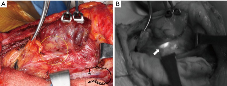
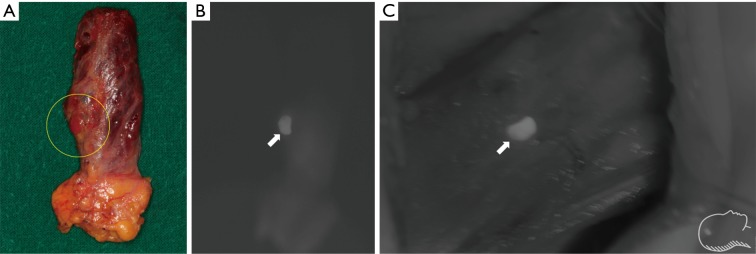
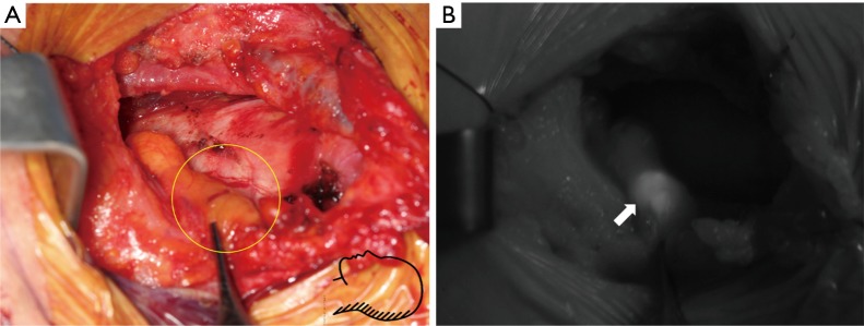
Surgeons have cited difficulties in identifying the parathyroid glands (PG) during thyroidectomy. To overcome the limitation of naked eye, many studies on near-infrared fluorescence imaging of PGs have been introduced and suggested that fluorescence imaging is useful for both localizing PGs and evaluating their function. This imaging technique has been reported in two ways: (I) imaging using a fluorescent material called indocyanine green (ICG); and (II) autofluorescence using intrinsic fluorophores. These innovative and novel techniques are expected to have a significant impact on performing thyroid or parathyroid surgery. In this article, current papers that describe ICG fluorescence and autofluorescence imaging of PG during thyroid and parathyroid surgery are reviewed.

Keywords: Parathyroid gland (PG); autofluorescence; near-infrared.


Conflict of interest statement

Conflicts of Interest: The authors have no conflicts of interest to declare.

Figures

Figure 1
Figure 1
Preparation of indocyanine green. ICG was mixed with 10 mL of sterile water, and 3–5 mL was administrated intravenously. The injection can be repeated until a maximum dose of 5 mg/kg/day is reached. This figure was kindly provided by courtesy of Professor Yoon Woo Koh, Department of Otolaryngology-Head and Neck Surgery of Yonsei University, Korea. ICG, indocyanine green.
Figure 2
Figure 2
Intraoperative NIR ICG images showing identification of normal parathyroid gland during retroauricular robotic thyroidectomy (DaVinci Si system, near infrared camera). (A) A right inferior parathyroid gland observed by naked eye under white light imaging (circle); (B) ahead of administration of ICG. The parathyroid gland was not clearly distinguishable with NIR mode imaging; (C) post-administration of ICG. The fluorescence was observed from the both parathyroid gland (arrow) and thyroid gland; (D) the intensity of the parathyroid gland (arrow) appeared to be higher. Distinction between the parathyroid gland and thyroid became clear as the parathyroid gland was separated from the thyroid gland. These figures were kindly provided by courtesy of Professor Yoon Woo Koh, Department of Otolaryngology-Head and Neck Surgery of Yonsei University, Korea. NIR, near-infrared; ICG, indocyanine green.
Figure 3
Figure 3
NIR ICG imaging of the parathyroid adenoma during retroauricular robotic thyroidectomy (DaVinci Si system, near infrared camera). (A) Photo taken with near infrared mode light before administrating ICG; (B) the image after ICG injection. Both parathyroid (circle) and thyroid gland were stained. The parathyroid gland (arrow) was dissected with naked eye (C) and localized with ICG fluorescence imaging (D). These figures were kindly provided by courtesy of Professor Yoon Woo Koh, Department of Otolaryngology-Head and Neck Surgery of Yonsei University, Korea. NIR, near-infrared; ICG, indocyanine green.
Figure 4
Figure 4
Images showing how to take a picture. (A) A photo of equipment setting in the operating room; (B) schematic diagram for detecting autofluorescence of parathyroid tissue with simultaneous illumination of the background tissues.
Figure 5
Figure 5
Images for detecting left inferior parathyroidectomy during conventional open thyroidectomy. (A) White light image after thyroid lobe lateralization. Inferior parathyroid gland is expected to be in the circle; (B) autofluorescence with NIR illuminator image. Autofluorescence emission (arrow) coincided with inferior parathyroid gland. Whole surgical field could be visualized by NIR illumination. NIR, near-infrared;
Figure 6
Figure 6
Images for detecting left inferior parathyroidectomy during conventional open thyroidectomy. (A) White light image of thyroidectomy and central neck dissection specimen. Detection of a removed parathyroid gland from the thyroidectomy specimen (circle); (B) the parathyroid gland showed autofluorescence (arrow) even when it was removed and devascularized; (C) preparation for autotransplantation of parathyroid gland (arrow) to right sternocleidomastoid muscle.
Figure 7
Figure 7
Images showing unusual location of parathyroid tissue. (A) A suspicious intrathymic parathyroid gland (circle); (B) localization of intrathymic parathyroid gland by NIR autofluorescence imaging (arrow).

References

    1. Hundahl SA, Fleming ID, Fremgen AM, et al. A National Cancer Data Base report on 53,856 cases of thyroid carcinoma treated in the US, 1985-1995. Cancer 1998;83:2638-48. 10.1002/(SICI)1097-0142(19981215)83:12<2638::AID-CNCR31>3.0.CO;2-1 - DOI - PubMed
    1. Suh YJ, Choi JY, Chai YJ, et al. Indocyanine green as a near-infrared fluorescent agent for identifying parathyroid glands during thyroid surgery in dogs. Surg Endosc 2015;29:2811-7. 10.1007/s00464-014-3971-2 - DOI - PubMed
    1. Lee NJ, Blakey JD, Bhuta S, et al. Unintentional parathyroidectomy during thyroidectomy. Laryngoscope 1999;109:1238-40. 10.1097/00005537-199908000-00010 - DOI - PubMed
    1. Lin DT, Patel SG, Shaha AR, et al. Incidence of inadvertent parathyroid removal during thyroidectomy. Laryngoscope 2002;112:608-11. 10.1097/00005537-200204000-00003 - DOI - PubMed
    1. Sasson AR, Pingpank JF, Jr, Wetherington RW, et al. Incidental parathyroidectomy during thyroid surgery does not cause transient symptomatic hypocalcemia. Arch Otolaryngol Head Neck Surg 2001;127:304-8. 10.1001/archotol.127.3.304 - DOI - PubMed

LinkOut - more resources