

Clinicopathologic Features of Membranous-Like Glomerulopathy With Masked IgG Kappa Deposits
- PMID: 29142932
- PMCID: PMC5678740
- DOI: 10.1016/j.ekir.2016.08.012
Clinicopathologic Features of Membranous-Like Glomerulopathy With Masked IgG Kappa Deposits
Abstract
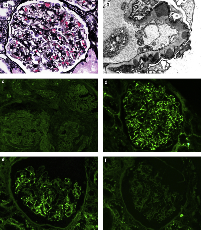
Introduction: Ig deposits identified on renal biopsy samples by paraffin immunofluorescence that show negative staining by routine immunofluorescence on frozen tissue have become known as "masked" deposits. Membranous-like glomerulopathy with masked IgG kappa (κ) deposits is a recently recognized pattern of immune complex deposition characterized by masked deposits that show IgG κ restriction and are subepithelial and mesangial by electron microscopy. Based on the frequent presence of C3-only staining by routine immunofluorescence microscopy (IF), these cases could be misdiagnosed as C3 glomerulonephritis in the absence of paraffin immunofluorescence evaluation.
Methods: The clinicopathologic details of all cases of membranous-like glomerulopathy with masked IgG κ deposits diagnosed in our laboratory were included, beginning with the initial recognition of this entity in 2011 through the end of 2015. Inclusion was based on renal biopsy sample morphologic features including glomerular deposits that stain for IgG κ and have a staining intensity that is significantly brighter by paraffin IF than by routine IF on frozen tissue.
Results: This pattern of immune complex deposition has been seen in 41 patients in our laboratory over a 5-year period. The patients with these biopsy findings are most commonly young female individuals with a mean age of 27.5 years, with 88% being less than 40 years. All patients had proteinuria with a mean 24-hour urine protein of 3.5 g (range 0.5-12.8 years) and 35% with nephrotic-range proteinuria. Hematuria was present in 88% of patients, and 29% had elevated serum creatinine at presentation. Autoimmune serologic tests were positive in 55% of patients, with a weakly positive antinuclear antibody being most common. Despite this, only 1 patient (2%) fulfilled the diagnostic criteria for systemic lupus erythematosus. The outcome data were mixed, as some patients showed spontaneous remission and mild disease whereas others progressed to end-stage renal disease. There was no apparent correlation between the treatment used and outcome in this retrospective analysis. One patient underwent transplantation and developed biopsy-proven recurrence of disease in the graft 42 months posttransplantation. The etiology of this entity remains unknown.
Discussion: We provide an expanded case series detailing the clinicopathologic findings of patients with membranous-like glomerulopathy with masked IgG κ deposits. Patients are most commonly young female individuals <40 years of age and commonly have positive autoimmune serologic studies such as antinuclear antibody, although few carry a diagnosis of any well-defined autoimmune disease such as lupus. The outcome data were mixed, as some patients showed spontaneous remission and mild disease whereas others progressed to ESRD. There was no apparent correlation between the treatment used and outcome in this retrospective analysis.
Keywords: C3 glomerulonephritis; masked deposits; membranous glomerulopathy; nephritis; proliferative glomerulonephritis with monoclonal IgG kappa deposits; renal biopsy.
Figures
References
-
- Glassock R.J. The pathogenesis of membranous nephropathy: evolution and revolution. Curr Opin Nephrol Hypertens. 2012;21:235–242. - PubMed
-
- Larsen C.P., Messias N.C., Silva F.G. Determination of primary versus secondary membranous glomerulopathy utilizing phospholipase A2 receptor (PLA2R1) staining in renal biopsies. Mod Pathol. 2013;26:709–715. - PubMed
-
- Larsen C.P., Ambruzs J.M., Bonsib S.M. Membranous-like glomerulopathy with masked IgG kappa deposits. Kidney Int. 2014;86:154–161. - PubMed
LinkOut - more resources
Full Text Sources
Other Literature Sources
Miscellaneous
