

doi: 10.1039/c7bm00872d.
Advanced smart-photosensitizers for more effective cancer treatment
Affiliations
- PMID: 29142997
- PMCID: PMC5736440
- DOI: 10.1039/c7bm00872d
Item in Clipboard
Advanced smart-photosensitizers for more effective cancer treatment
Biomater Sci.
.
Abstract
Photodynamic therapy (PDT) based upon the use of light and photosensitizers (PSs) has been used as a novel treatment approach for a variety of tumors. It, however, has several major limitations in the clinic: poor water solubility, long-term phototoxicity, low tumor targeting efficacy, and limited light penetration. With advances in nanotechnology, materials science, and clinical interventional imaging procedures, various smart-PSs have been developed for improving their cancer-therapeutic efficacy while reducing the adverse effects. Here, we briefly review state-of-the-art smart-PSs and discuss the future directions of PDT technology.
Figures

(a–b) (a) Chemical structure of the T-PPS and (b) their bioapplications. (c–d) Temperature-dependent change in (c) transmittance of T-PPS and native HPC (inset: photograph of T-PPS aqueous solution (10 mg/mL in water) at (1) T < lower critical solution temperature (LCST) and (2) T > LCST), and (d) singlet oxygen generation of T-PPS in aqueous solution (n = 3, *p < 0.001). (e–f) In vitro photocytotoxicity of T-PPS at various temperatures on PANC-2 cells. (e) Cell counting based upon kit-8 (CCK-8) assay and (f) Live/dead assay for cell viability of PANC-2 cells incubated with T-PPS (concentration of PPb-a, 1 μg/mL) under laser irradiation (3.0 J/cm2) at 37 °C and 45 °C (n = 4, *p < 0.002). Reproduced with permission from ref. . Copyright 2016 American Chemical Society.

(a) Schematic illustration of luminescence and magnetic resonance (MR) imaging with photodynamic therapy (PDT) using UCNP-based PS. (b) Transmission electron microscopy (TEM) image of NaYF4:Yb,Er/NaGdF4 core–shell UCNP. (c) Bright field and upconversion luminescence images of a nude mouse bearing tumor after intravenous injection of UCNP-based PS. (d) T1-weighted color maps of a tumor-bearing nude mouse before and 1.5 h after intravenous injection of UCNP-based PS. Arrows indicate tumor sites. (e) Growth of tumors after treatments (n=3 for each group). Reproduced with permission from ref. . Copyright 2012 Wiley-VCH Verlag GmbH & Co. KGaA, Weinheim.

(a) Chemical structure of polydopamine nanoparticle (PD-NP) and PS-conjugated hyaluronic acid (PS-HA) conjugates. (b) Schematic illustration of synergistic cancer therapy effect of PDT and PTT using PS–hyaluronic acid conjugates shielded polydopamine nanoparticle (PHPD-NP). (c) Scanning electron microscopy (SEM) image of PHPD-NP (scale bar: 100 nm). (d) Live/Dead assay of MDA-MB-231 cells treated with PHPD-NP with or without laser irradiation at 3.0 J/cm2 power. (e) Tumor growth inhibition after intratumoral injection of phosphate-buffered saline (PBS), PD-NP, PS-HAs, or PHPD-NPs with laser irradiation (n = 4). Reproduced with permission from ref. . Copyright 2016 American Chemical Society.
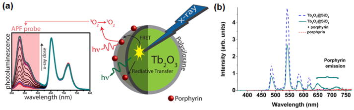
(a) Schematic illustration of the working mechanism of X-PDT. Under X-ray irradiation, nanoscintillator (Tb2O3) transfer energy toward the photosensitizer (PS, Porphyrin), leading to singlet oxygen generation. (b) Fluorescence spectra measurement performed with 300 nm excitation. The emission spectrum of porphyrin was almost zero, while the spectrum of the porphyrin grafted Tb2O3@SiO3 nanoparticle (NP) presents both the emission peaks at 650 and 725 nm. Reproduced with permission from ref. . Copyright 2013 American Chemical Society.
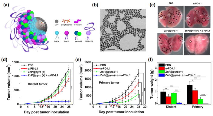
(a) Scheme illustration of the Zn-pyrophosphate (ZnP) core and the photosensitizer (PS) pyrolipid (a lipid conjugate of pyropheophorbide-a) shell of the immunogenic NP (ZnP@pyro). (B) Transmission electron microscopy (TEM) image showing the spherical and nearly monodispersed morphology of ZnP@pyro (scale bar = 200 nm). (c) Photographs of excised primary 4T1 tumors at the end point (23 days after tumor implantation). From top to bottom: phosphate-buffered saline (PBS), α-PD-L1, ZnP@pyro (+), and ZnP@pyro (+) plus α-PD-L1. The tumors indicated with dot-lined rectangle disappeared in the ZnP@pyro (+) + α-PD-L1 group. This result suggests that the ZnP@pyro PDT combined with PD-L1 blockade eradicates primary 4T1 tumors and prevents lung metastasis. (d–e) (d) Distant and (e) primary tumor growth curves of 4T1 models. The arrows represent the time of NP administration (black) and irradiation (red). (f) Weight of 4T1 tumors at the end point (28 days after tumor implantation) of the experiment. “(+)” in the figure legends refers to treatment with irradiation. *P < 0.05, ** P < 0.01, *** P < 0.001. The combination of ZnP@pyro with irradiation and PD-L1 blockade induced complete eradication of the irradiated primary tumors (synergistic effect) and effective control of the nonirradiated distant tumors (abscopal effect), eliciting a 92% reduction in tumor size compared to the PBS control group. These results indicate that tumors can be sensitized to PD-L1 blockade immunotherapy by ZnP@pyro PDT-mediated tumor-specific immune responses. Reproduced with permission from ref. . Copyright 2016 American Chemical Society.

(a) The mechanism of action of PDT is illustrated by the Jablonski diagram. When light (photon) is shone on PS, the PS can be excited to the first excited singlet state that can relax to the more long-lived triplet state. The triplet PS can interact with oxygen molecules to produce reactive oxygen species (ROS) through the type 1 and type 2 mechanisms. (b) Classification of the generations of PSs developed so far. Currently developed PSs can be divided into first generation and second-generation based upon their physico-chemical property. The third-generation of smart PSs functionalized with polymers or nanoparticle is currently under development.
References
MeSH terms
Substances
Grants and funding
LinkOut - more resources
Full Text Sources
Other Literature Sources