. 2018 Feb 1;596(3):393-407.
doi: 10.1113/JP275178. Epub 2017 Dec 18.

K2P TASK-2 and KCNQ1-KCNE3 K+ channels are major players contributing to intestinal anion and fluid secretion

Affiliations

K2P TASK-2 and KCNQ1-KCNE3 K+ channels are major players contributing to intestinal anion and fluid secretion

Francisca Julio-Kalajzić et al. J Physiol. .

Abstract

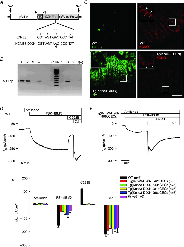
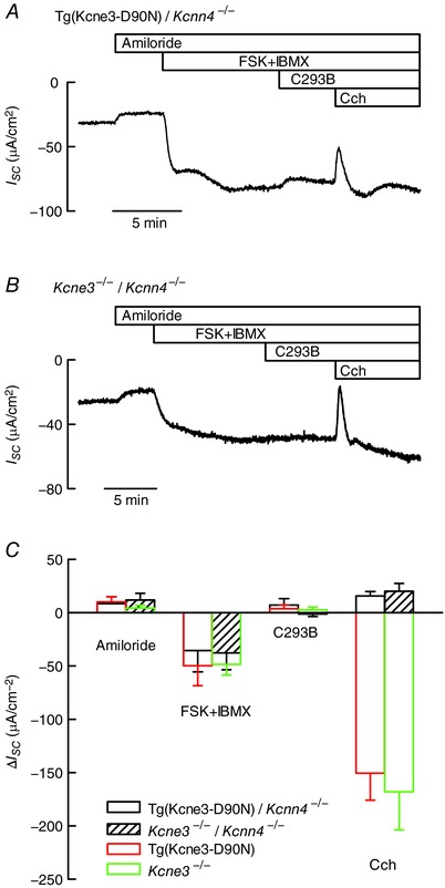
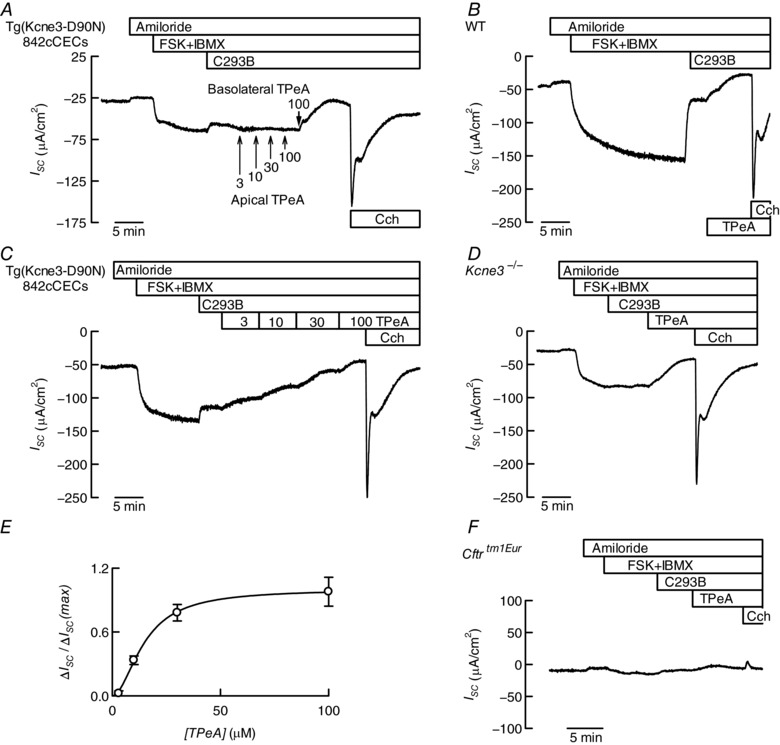
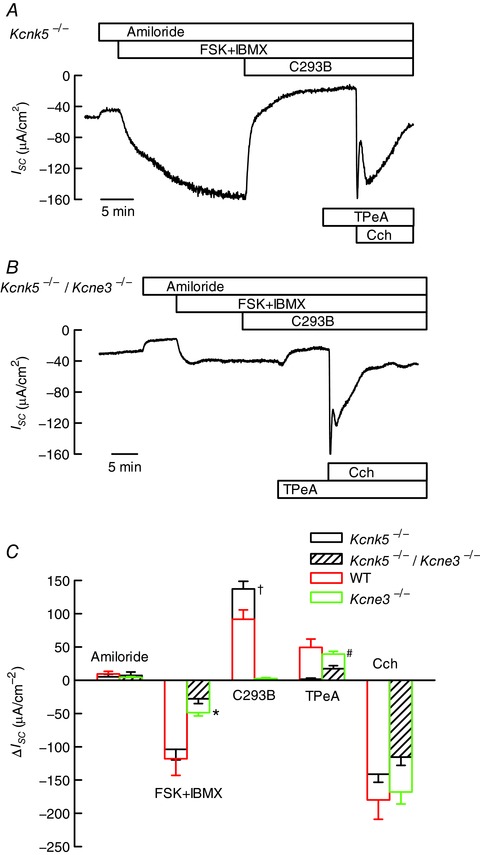
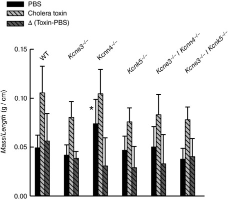
Key points: K+ channels are important in intestinal epithelium as they ensure the ionic homeostasis and electrical potential of epithelial cells during anion and fluid secretion. Intestinal epithelium cAMP-activated anion secretion depends on the activity of the (also cAMP dependent) KCNQ1-KCNE3 K+ channel, but the secretory process survives after genetic inactivation of the K+ channel in the mouse. Here we use double mutant mice to investigate which alternative K+ channels come into action to compensate for the absence of KCNQ1-KCNE3 K+ channels. Our data establish that whilst Ca2+ -activated KCa 3.1 channels are not involved, K2P two-pore domain TASK-2 K+ channels are major players providing an alternative conductance to sustain the intestinal secretory process. Work with double mutant mice lacking both TASK-2 and KCNQ1-KCNE3 channels nevertheless points to yet-unidentified K+ channels that contribute to the robustness of the cAMP-activated anion secretion process.

Abstract: Anion and fluid secretion across the intestinal epithelium, a process altered in cystic fibrosis and secretory diarrhoea, is mediated by cAMP-activated CFTR Cl- channels and requires the simultaneous activity of basolateral K+ channels to maintain cellular ionic homeostasis and membrane potential. This function is fulfilled by the cAMP-activated K+ channel formed by the association of pore-forming KCNQ1 with its obligatory KCNE3 β-subunit. Studies using mice show sizeable cAMP-activated intestinal anion secretion in the absence of either KCNQ1 or KCNE3 suggesting that an alternative K+ conductance must compensate for the loss of KCNQ1-KCNE3 activity. We used double mutant mouse and pharmacological approaches to identify such a conductance. Ca2+ -dependent anion secretion can also be supported by Ca2+ -dependent KCa 3.1 channels after independent CFTR activation, but cAMP-dependent anion secretion is not further decreased in the combined absence of KCa 3.1 and KCNQ1-KCNE3 K+ channel activity. We show that the K2P K+ channel TASK-2 is expressed in the epithelium of the small and large intestine. Tetrapentylammonium, a TASK-2 inhibitor, abolishes anion secretory current remaining in the absence of KCNQ1-KCNE3 activity. A double mutant mouse lacking both KCNQ1-KCNE3 and TASK-2 showed a much reduced cAMP-mediated anion secretion compared to that observed in the single KCNQ1-KCNE3 deficient mouse. We conclude that KCNQ1-KCNE3 and TASK-2 play major roles in the intestinal anion and fluid secretory phenotype. The persistence of an, admittedly reduced, secretory activity in the absence of these two conductances suggests that further additional K+ channel(s) as yet unidentified contribute to the robustness of the intestinal anion secretory process.

Keywords: K+ channel; epithelial transport; fluid secretion.


Figures

Figure 1
Figure 1. Generation and characterization of transgenic mice expressing KCNE3‐D90N mutant in intestinal epithelium
A, scheme of the pVillin KCNE3‐D90N‐HA transgene used for the generation of tissue‐specific transgenic mice expressing hemagglutinin (HA)‐tagged KCNE3‐D90N protein under intestinal epithelium‐specific promoter villin; SV40(A), simian virus 40 polyadenylation signal. SalI, restriction site used for transgene (9600 bp) linearization. The arrowheads indicate the primer binding sites. Bottom, sequence of KCNE3, showing the G>A mutation results in KCNE3‐D90N protein. B, a 560 bp PCR fragment is detectable with the P1–P2 primer pair in transgenic mice. Lanes labelled 1, 2, 4 and 8 correspond to different KCNE3‐D90N transgenic lines (later named as in panel F) and lanes 3, 5, 6, 7 and 9 to mice that did not incorporate the transgene; negative control C (−) is DNA from a WT biopsy. C, tissue distribution of KCNE3‐D90N and KCNQ1. KCNE3‐D90N was detected using immunofluorescence against HA (green) in mouse distal colon. Top panels show WT control sections; bottom, transgenic tissues from the Tg(Kcne3‐D90N)842cCECs line. Right, staining for KCNQ1 (red). Insets show higher magnifications of regions shown in frames with arrowheads pointing to basolateral aspect of enterocytes. Bar represents 40 μm. D and E, traces showing recordings of short circuit currents (I SC) as function of time obtained using distal colon from WT (D) or Tg(Kcne3‐D90N) (E) mice mounted in Ussing chambers. Additions, during the times shown in the upper bars, were 10 μm amiloride, 100 μm isobutylmethylxanthine (IBMX) plus 1 μm forskolin (FSK), 10 μm chromanol 293B (C293B) and 100 μm carbachol (Cch). All additions were made to the serosal side of the epithelium save for amiloride, which was added apically. Tissue resistances were, respectively, 78 and 71 Ω cm2 at the beginning and end of the experiment shown in D. The respective numbers in E were 91 and 67 Ω cm2. F, bar graph summarizing the mean change in short circuit‐current (ΔI SC) after the different additions in D and E. Each colour corresponds to a different Tg(Kcne3‐D90N) transgenic line. Data for the Kcne3 −/− mouse are also shown. Results are means ± SD of the number of animals given in parentheses. FSK+IBMX and C293B treatments in transgenic and Kcne3 −/− tissues are all different from those in WT judged by Bonferroni t test with P < 0.001.
Figure 2
Figure 2. Effect of additional inactivation of the Ca2+‐dependent K+ channel KCa3.1 on anion secretion in the colon of Kcne3 null animals
The double mutants were obtained by crossing single mutants expressing a dominant‐negative KCNE3‐D90N subunit or Kcne3 −/− mice with KCa3.1 knockouts (Kcnn4 −/−). A and B, traces of short circuit currents (I SC) as function of time obtained using mouse distal colon of Tg(KCNE3‐D90N)/Kcnn4 −/− or Kcne3 −/−/Kcnn4 −/− homozygous double mutants mounted in Ussing chambers. Details of compound additions are as in Fig. 1. Tissue resistances were, respectively, 68 and 60 Ω cm2 at the beginning and end of the experiment shown in A. The respective values in B were 78 and 67 Ω cm2. C, summary of the results of experiments in A and B. Data are means ± SD (number of animals: 5 for Tg(KCNE3‐D90N)/Kcnn4 −/− and 6 for Kcne3 −/−/Kcnn4 −/−). The results for single Tg(Kcne3‐D90N) transgenic and Kcne3 −/− shown in Fig. 1 F are reproduced for comparison. The transgenic line used in these experiments was Tg(Kcne3‐D90N)843cCECs. There was no difference between the results obtained using single or double mutants, except for the complete absence of carbachol‐induced anion secretion in the tissues of animals lacking KCa3.1.
Figure 3
Figure 3. Inhibiting cAMP‐dependent intestinal anion secretion by pharmacological K+ channel blockade
AD and F, Ussing chamber traces of short circuit currents (I SC) as function of time obtained using distal colon of transgenic mice expressing the KCNE3‐D90N mutant subunit (A and C), a WT mouse (B), a Kcne3 −/− (D) and a Cftrtm1Eur mouse expressing the CFTR‐ΔF508 mutation (F). Details of compound additions are as in Fig. 1 except for the use of tetrapentylammonium (TPeA) added, unless otherwise indicated, basolaterally at 100 μm. Tissue resistances at the beginning and end of the experiment were 82 and 68 Ω cm2 in A, 98 and 70 Ω cm2 in B, 66 and 58 Ω cm2 in D, and 82 and 82 Ω cm2 in F. E, concentration dependence of the effect of basolateral TPeA on the cAMP‐activated anion secretion (normalized) measured using distal colon from Tg(Kcne3‐D90N)842cCECs mice expressing the KCNE3‐D90N subunit (means ± SD, n = 4).
Figure 4
Figure 4. Expression of TASK‐2 in the intestinal epithelium of the colon revealed by β‐galactosidase activity in Kcnk5 −/− mice
Ileum (A, B, E and F) or colon (C, D, G and H) sections from Kcnk5 −/− (A, C, E and G) and WT (B, D, F and H) mice were stained for X‐gal (blue) thus revealing β‐galactosidase activity used here as a proxy for endogenous Kcnk5 gene expression. Bars: 500 μm for the four upper panels and 100 μm for the lower panels.
Figure 5
Figure 5. Anion secretion in the colon of mice deficient in K2P K+ channel TASK‐2 or in double mutants lacking expression of both TASK‐2 and KCNQ1–KCNE3 K+ channels
The double mutants were obtained by crossing single mutant mice. A and B, traces of short circuit currents (I SC) as function of time obtained using mouse distal colon of Kcnk5 −/− or Kcnk5 −/−/Kcne3 −/− homozygous double mutants mounted in Ussing chambers. Details of compound additions are as in Figs 1 and 3. Tissue resistances were, respectively, 81 and 66 Ω cm2 at the beginning and end of the experiment shown in A. The respective numbers in B were 102 and 71 Ω cm2. C, summary of the results of experiments in A and B. Data are means ± SD (number of animals: 6 for Kcnk5 −/− and 6 for Kcnk5 −/−/Kcne3 −/−). The results for WT and single Kcne3 −/− from Fig. 1 are reproduced for comparison. Results for double Kcnk5 −/−/Kcne3 −/− and single Kcne3 −/− mutants were different when tested by t test as indicated: * P < 0.01; † and #, P < 0.001.
Figure 6
Figure 6. Cholera toxin‐induced small intestinal fluid secretion measured in vivo
Mass over length relations of ileal loops 5 h after luminal injection of PBS or PBS + cholera toxin (0.5 μg per loop) in wild‐type (WT) and various single and double knockout mouse models. The difference between the measurements taken without the toxin was subtracted from the toxin‐containing number to give the net secretion stimulated by cholera toxin (Δ (Toxin‐PBS)). Data are means ± SD. The numbers of experiments were as follows: WT, n = 10; Kcne3 −/−, n = 10; Kcnn4 −/−, n = 9; Kcnk5 −/−, n = 4; Kcnn4 −/−/Kcne3 −/−, n = 11; Kcnk5 −/−/Kcne3 −/−, n = 10. There was no difference between measurements on mutant mice and that obtained with WT except for measurement with PBS alone identified with *(P = 0.002 by ANOVA).

Comment in

Similar articles

Cited by

References

    1. Abbott GW & Goldstein SA (2002). Disease‐associated mutations in KCNE potassium channel subunits (MiRPs) reveal promiscuous disruption of multiple currents and conservation of mechanism. FASEB J 16, 390–400. - PubMed
    1. Ahuja M, Jha A, Maleth J, Park S & Muallem S (2014). cAMP and Ca2+ signaling in secretory epithelia: crosstalk and synergism. Cell Calcium 55, 385–393. - PMC - PubMed
    1. Al‐Hazza A, Linley J, Aziz Q, Hunter M & Sandle G (2016). Upregulation of basolateral small conductance potassium channels (KCNQ1/KCNE3) in ulcerative colitis. Biochem Biophys Res Commun 470, 473–478. - PMC - PubMed
    1. Al‐Hazza A, Linley JE, Aziz Q, Maclennan KA, Hunter M & Sandle GI (2012). Potential role of reduced basolateral potassium (IKCa3.1) channel expression in the pathogenesis of diarrhoea in ulcerative colitis. J Pathol 226, 463–470. - PubMed
    1. Alzamora R, O'Mahony F, Ko WH, Yip TW, Carter D, Irnaten M & Harvey BJ (2011). Berberine reduces cAMP‐induced chloride secretion in T84 human colonic carcinoma cells through inhibition of basolateral KCNQ1 channels. Front Physiol 2, 33. - PMC - PubMed

Publication types

LinkOut - more resources