

Spontaneous neural activity differences in posttraumatic stress disorder: A quantitative resting-state meta-analysis and fMRI validation
- PMID: 29143411
- PMCID: PMC6866285
- DOI: 10.1002/hbm.23886
Spontaneous neural activity differences in posttraumatic stress disorder: A quantitative resting-state meta-analysis and fMRI validation
Abstract
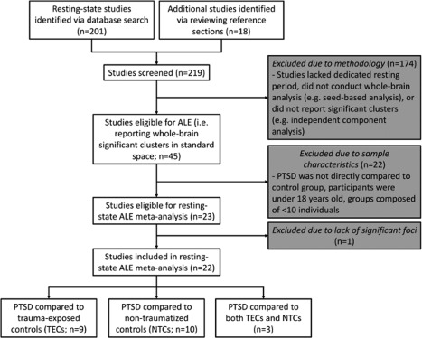
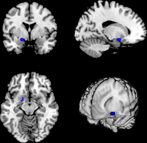
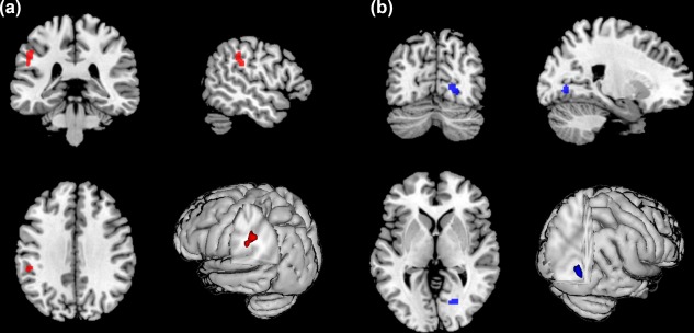
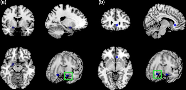
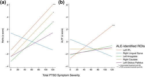
Identifying the pathophysiology of posttraumatic stress disorder (PTSD) is a critical step toward reducing its debilitating impact. Spontaneous neural activity, measured at rest using various neuroimaging techniques (e.g., regional homogeneity [ReHo], amplitude of low frequency fluctuations [ALFF]), can provide insight about baseline neurobiological factors influencing sensory, cognitive, or behavioral processes associated with PTSD. The present study used activation likelihood estimation (ALE) to conduct the largest-to-date quantitative meta-analysis of spontaneous neural activity in PTSD, including 457 PTSD cases, 292 trauma-exposed controls (TECs), and 293 non-traumatized controls (NTCs) across 22 published studies. Five regions-of-interest (ROIs) were identified where activity differed between PTSD cases and controls: one when compared to all controls (left globus pallidus), two when compared to TECs (left inferior parietal lobule [IPL] and right lingual gyrus), and two when compared to NTCs (left amygdala and right caudate head). To corroborate these results, a second analysis was conducted using resting-state functional magnetic resonance imaging on an independent sample of 205 previously-deployed US military veterans. In this analysis, converging evidence from ReHo and ALFF showed that spontaneous neural activity in the left IPL alone was positively correlated with PTSD symptom severity. This result is consistent with theoretical accounts that link left IPL activity with PTSD-relevant processes such as processing of emotional stimuli (e.g., fearful faces) and the extent that attention is captured by salient autobiographical memories. By modeling the neurobiological correlates of PTSD, we can increase our understanding of this debilitating disorder and guide the development of future clinical innovations.
Keywords: amygdala; globus pallidus; magnetic resonance imaging; neuroimaging; occipital lobe; parietal lobe; post-traumatic stress disorder; rest; veterans.
© 2017 Wiley Periodicals, Inc.
Figures
References
-
- Aiello, M. , Salvatore, E. , Cachia, A. , Pappatà, S. , Cavaliere, C. , Prinster, A. , … Quarantelli, M. (2015). Relationship between simultaneously acquired resting‐state regional cerebral glucose metabolism and functional MRI: a PET/MR hybrid scanner study. Neuroimage, 113, 111–121. 10.1016/j.neuroimage.2015.03.017 - DOI - PubMed
-
- American Psychiatric Association . (2000). Diagnostic criteria from DSM‐IV‐TR. Arlington, VA: Am Psychiatric Association.
-
- American Psychiatric Association . (2013). Diagnostic and statistical manual of mental disorders. American Psychiatric Publishing, Incorporated.
-
- Andersson, J. L. R. , Skare, S. , & Ashburner, J. (2003). How to correct susceptibility distortions in spin‐echo echo‐planar images: application to diffusion tensor imaging. Neuroimage, 20(2), 870–888. - PubMed
Publication types
MeSH terms
Grants and funding
LinkOut - more resources
Full Text Sources
Other Literature Sources
Medical
