. 2018;17(1):110-123.
doi: 10.1080/15384101.2017.1404207. Epub 2018 Jan 8.

Mammalian target of rapamycin complex 2 (mTORC2) controls glycolytic gene expression by regulating Histone H3 Lysine 56 acetylation

Affiliations

Mammalian target of rapamycin complex 2 (mTORC2) controls glycolytic gene expression by regulating Histone H3 Lysine 56 acetylation

Raghavendra Vadla et al. Cell Cycle. 2018.

Abstract

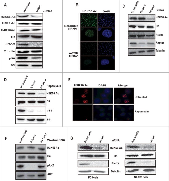
Metabolic reprogramming is a hallmark of cancer cells, but the mechanisms are not well understood. The mammalian target of rapamycin complex 2 (mTORC2) controls cell growth and proliferation and plays a critical role in metabolic reprogramming in glioma. mTORC2 regulates cellular processes such as cell survival, metabolism, and proliferation by phosphorylation of AGC kinases. Components of mTORC2 are shown to localize to the nucleus, but whether mTORC2 modulates epigenetic modifications to regulate gene expression is not known. Here, we identified histone H3 lysine 56 acetylation (H3K56Ac) is regulated by mTORC2 and show that global H3K56Ac levels were downregulated on mTORC2 knockdown but not on mTORC1 knockdown. mTORC2 promotes H3K56Ac in a tuberous sclerosis complex 1/2 (TSC1/2) mediated signaling pathway. We show that knockdown of sirtuin6 (SIRT6) prevented H3K56 deacetylation in mTORC2 depleted cells. Using glioma model consisting of U87EGFRvIII cells, we established that mTORC2 promotes H3K56Ac in glioma. Finally, we show that mTORC2 regulates the expression of glycolytic genes by regulating H3K56Ac levels at the promoters of these genes in glioma cells and depletion of mTOR leads to increased recruitment of SIRT6 to these promoters. Collectively, these results identify mTORC2 signaling pathway positively promotes H3K56Ac through which it may mediate metabolic reprogramming in glioma.

Keywords: Cancer cells; EGFR; Glioma; Histone acetylation; Histone deacetylases; SIRT6; TSC1/2; growth factors; mTOR; metabolic reprogramming; metabolism; signal transduction.


Figures

Figure 1.
Figure 1.
mTORC2 regulates H3K56 acetylation. (A) mTOR signaling positively regulates global H3K56Ac levels but not H3K9Ac and H4K16Ac. HeLa cells were transfected with scramble or mTOR siRNA using lipofectamine reagent. After 48 h of transfection, cells were harvested, whole cell lysates were prepared and resolved on SDS-PAGE and levels of indicated proteins were analyzed by Western blot. (B) HeLa cells were transfected with scramble or mTOR siRNA. After 48 h of transfection, cells were fixed with paraformaldehyde. Immunofluorescence was performed using anti-H3K56Ac antibodies. Images shown are from a representative of multiple experiments. (C) Rictor but not raptor knockdown downregulates H3K56Ac. HeLa cells were transfected with raptor or rictor siRNA. After 48 h cells were harvested and whole cell lysates were prepared. Lysates were resolved on SDS-PAGE and the levels of H3K56Ac was analyzed by Western blot. (D) Prolonged treatment with rapamycin downregulates H3K56Ac. HeLa cells were treated with 100nM rapamycin for 2 and 24 h, whole cell lysates were prepared and equal quantities of cell lysates were resolved on SDS-PAGE and the levels of H3K56Ac was analysed by Western blot. (E) HeLa cells were treated with rapamycin (100nM) for 24 h. Cells without treatment were taken as controls. After 24 h treatment, cells were fixed with paraformaldehyde and immunofluorescence was performed with anti-H3K56Ac antibodies. (F) H3K56Ac levels were unchanged on Wortmannin treatment. HeLa cells were kept untreated or treated with wortmannin (100nM) for 2 h or 24 h and harvested. Whole cell lysates were resolved on SDS-PAGE and the levels of indicated proteins were analyzed by Western blot. (G) mTORC2 mediated regulation of H3K56Ac is conserved in different cell lines. PC3 and NIH3T3 cells were transfected with scramble or rictor siRNA. After 48 h of transfection, cells were harvested and whole cell lysates were prepared, resolved on SDS-PAGE. Levels of H3K56Ac were analyzed by western blot. Total H3 and Tubulin were used as loading controls.
Figure 2.
Figure 2.
TSC complex functions upstream of mTORC2 to regulate H3K56 acetylation. (A) TSC complex positively regulates H3K56Ac. HeLa cells were transfected with scramble, TSC1 or TSC2 siRNA. After 48 h of transfection, cells were harvested. Whole cell lysates were prepared and resolved on SDS-PAGE and levels of H3K56Ac were analyzed by Western blot. (B) mTORC2 mediated AKTser473 phosphorylation is downregulated in TSC2 depleted cells. HeLa cells were transfected with TSC2 siRNA. After 48 h, cells were harvested and whole cell lysates were analysed for Aktser473 phosphorylation by Western blot. AKT was probed as a loading control. (C) TSC2 regulate H3K56Ac by both mTORC2 dependent and independent pathways. HeLa cells were transfected with either scramble, TSC2 or rictor siRNA alone or transfected with both TSC2 and rictor siRNA. After 48 h of transfection, cells were harvested and whole cell lysates were loaded on SDS-PAGE and analyzed for H3K56Ac by Western blot. (D) Overexpression of rictor and TSC2 proteins induce H3K56Ac. HeLa cells were transfected with flag-rictor or pcDNA3.1-TSC2 DNA constructs for 24 h. Whole cell lysates were prepared and resolved on SDS-PAGE. Levels of H3K56Ac was analysed by Western blot. H3 and Tubulin were probed as loading controls. (E) Overexpression of TSC2N1643S (GAP mutant) induce H3K56Ac. HeLa cells were transfected with TSC2 or TSC2N1643S DNA constructs for 24h. Whole cell lysates were analyzed for the levels of H3K56Ac by Western blot.
Figure 3.
Figure 3.
SIRT6 deacetylates H3K56Ac in the absence of mTORC2. (A) Disruption of mTORC2 signaling does not alter SIRT6 expression. HeLa cells were transfected with scramble, rictor, or TSC2 siRNA. After 48 h of transfection, cells were harvested and whole cell lysates were resolved on SDS-PAGE. Levels of SIRT6 were analyzed by Western blot. (B) SIRT6 deacetylates H3K56 in the absence of mTOR. HeLa cells were transfected with scramble, mTOR or in a combination of mTOR and SIRT6 siRNA. After 72 h of transfection, whole cell lysates were analyzed for H3K56Ac levels by Western blot. (C) SIRT6 deacetylates H3K56 in the absence of TSC complex. HeLa cells were transfected with either scramble, TSC1, TSC2 siRNA or in combination with SIRT6 siRNA. After 48 h of transfection, whole cell lysates were analyzed for H3K56Ac levels by Western blot. (D) The decrease in H3K56Ac resulting from rictor depletion is recovered by SIRT6 knockdown. HeLa cells were transfected with either scramble, rictor siRNA or in combination with SIRT6 siRNA. After 48 h of transfection, whole cell lysates were analysed for H3K56Ac levels by Western blot. (E) Decrease in AKT S473 phosphorylation resulting from rictor depletion cannot be rescued by SIRT6 knockdown. HeLa cells were transfected with scramble or rictor siRNA alone or transfected with rictor and SIRT6 siRNA. After 48 h of transfection, whole cell lysates were analyzed for Aktser473 phosphorylation and AKT levels by Western blot. (F) mTORC2 negatively regulate deacetylase activity of SIRT6. HeLa cells were transfected with scramble or rictor siRNA for 48 h and then cells were transfected with the FLAG-SIRT6 plasmid for 24 hours. Whole cell lysates were resolved on SDS-PAGE and analyzed for indicated proteins by Western blot. (G) SIRT6 interacts with rictor. Endogenous SIRT6 was co-immunoprecipitated from HeLa cells with SIRT6 specific antibody and lysates were analyzed for the presence of rictor by Western blot.
Figure 4.
Figure 4.
mTORC2 signaling upregulates H3K56Ac in glioma. (A) EGF upregulates global H3K56Ac. HeLa cells were serum starved overnight followed by treatment with EGF (100 ng/ml) for 10 min, whole cell lysates were analyzed for levels of H3K56Ac by Western blot. Quantitative bar graph demonstrates relative H3K56Ac levels. Data are represented as mean ± SD of three independent experiments. Significant P-values were obtained with Student's t-test. ***P<0.001. (B) Knockdown of rictor abrogates EGF induced H3K56Ac in HeLa cells. HeLa cells were transfected with scramble or rictor siRNA. After 48 h, cells were serum starved overnight followed by treatment with EGF (100 ng/ml) for 10 min. Cells were harvested and whole cell lysates were analyzed for H3K56Ac by Western blot. (C) H3K56Ac levels were increased in U87EGFRvIII cells. Levels of H3K56Ac was analyzed in whole cell lysates of U87 and U87 EGFRVIII cells by western blot. (D) Knockdown of rictor downregulates H3K56Ac in U87EGFRVIII cells. U87EGFRvIII cells were transfected with scramble or rictor siRNA. After 72 h of transfection, whole cell lysates were analyzed for H3K56Ac by Western blot. (E) U87EGFRVIII cells were transfected with either scramble or rictor siRNA alone or both rictor and SIRT6 siRNA. At 72 h post transfection, whole cell lysates were analyzed for levels of H3K56Ac by Western blot. (F) overexpression of rictor in U87 cells induces H3K56Ac. U87 cells were transfected with empty vector or Flag-rictor DNA constructs and H3K56Ac was analyzed in whole cell lysates by Western blot.
Figure 5.
Figure 5.
mTORC2 and SIRT6 regulates expression of glycolytic genes in glioma. Rictor and SIRT6 regulate glycolytic gene expression in U87EGFRVIII cells. U87EGFRvIII cells were transfected with scramble or rictor or SIRT6 or both rictor and SIRT6 siRNA. At 48 h post transfection, cells were harvested. RNA was isolated by using Trizol method and total RNA was used for cDNA synthesis using Superscript III reverse transcriptase. mRNA levels of PDK1, LDHB, GLUT1 and LDHA genes were analyzed by RT-PCR. Data represents mean ± SEM from three independent experiments and was analyzed using a two-tailed unpaired Student's t-test. **P ≤ 0.01; *P ≤ 0.05.
Figure 6.
Figure 6.
mTORC2 regulates expression of glycolytic genes by modulating H3K56Ac at their promoters in glioma. (A) H3K56Ac decreases on promoters of PDK1, LDHB, GLUT1 and LDHA genes in the absence of rictor. U87EGFRvIII cells were transfected with scramble, rictor or both rictor and SIRT6 siRNA. At 48 h post transfection, cells were harvested. Chromatin immunoprecipitation (ChIP) was performed using H3K56Ac antibody and primers to the promoters of PDK1, LDHB, GLUT1 or LDHA genes. (B) Localisation of SIRT6 increases on the promoters of PDK1, LDHB, GLUT1 and LDHA genes in the absence of rictor. U87EGFRvIII cells were transfected with scramble, rictor or both rictor and SIRT6 siRNA. At 48 h post transfection, cells were harvested. Chromatin immunoprecipitation (ChIP) was performed using SIRT6 antibody and primers to the promoters of PDK1, LDHB, GLUT1 or LDHA genes.
Figure 7.
Figure 7.
Model depicting the novel function of mTORC2 signaling in promoting H3K56Ac by regulating localization of SIRT6 on the promoters of glycolytic genes resulting in their elevated expression in glioma.

References

    1. Laplante M, Sabatini DM. mTOR signaling in growth control and disease. Cell. 2012;149:274–293. doi: 10.1016/j.cell.2012.03.017. PMID:22500797 - DOI - PMC - PubMed
    1. Laplante M, Sabatini DM. mTOR signaling at a glance. J Cell Sci. 2009;122:3589–3594. doi: 10.1242/jcs.051011. PMID:19812304 - DOI - PMC - PubMed
    1. Frias MA, Thoreen CC, Jaffe JD, et al.. mSin1 is necessary for Akt/PKB phosphorylation, and its isoforms define three distinct mTORC2s. Curr Biol. 2006;16:1865–1870. doi: 10.1016/j.cub.2006.08.001. PMID:16919458 - DOI - PubMed
    1. Sarbassov DD, Guertin DA, Ali SM, et al.. Phosphorylation and regulation of Akt/PKB by the rictor-mTOR complex. Science. 2005;307:1098–1101. doi: 10.1126/science.1106148. PMID:15718470 - DOI - PubMed
    1. Yang Q, Inoki K, Ikenoue T, et al.. Identification of Sin1 as an essential TORC2 component required for complex formation and kinase activity. Genes Dev. 2006;20:2820–2832. doi: 10.1101/gad.1461206. PMID:17043309 - DOI - PMC - PubMed

Publication types