Review
. 2017 Oct 28;18(11):2272.
doi: 10.3390/ijms18112272.

Primary Cilium-Dependent Signaling Mechanisms

Affiliations
Review

Primary Cilium-Dependent Signaling Mechanisms

Rajasekharreddy Pala et al. Int J Mol Sci. .

Abstract

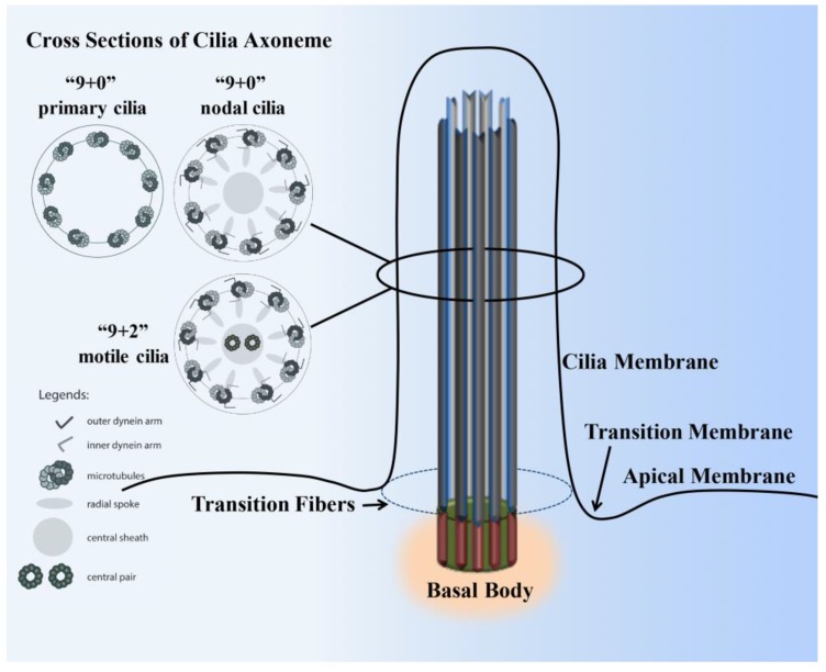
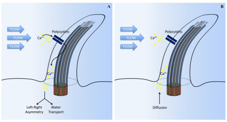
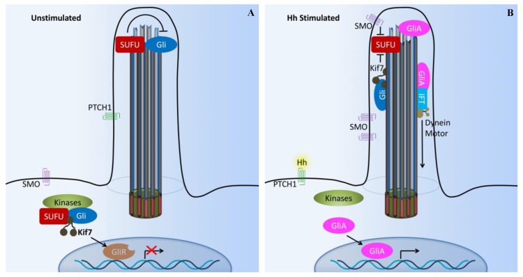
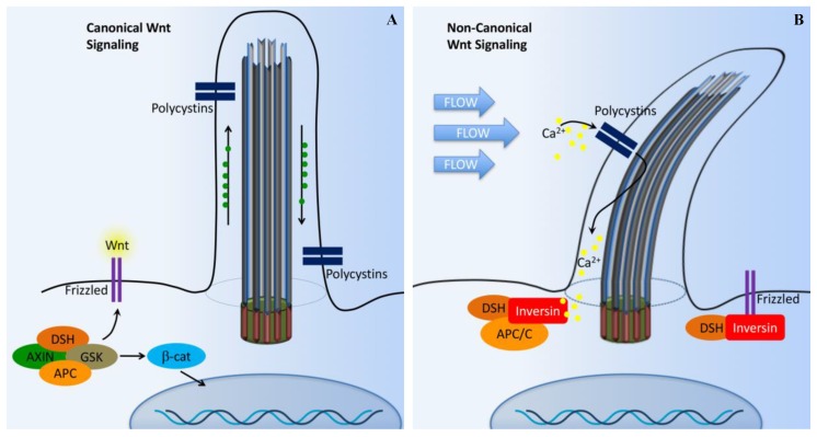
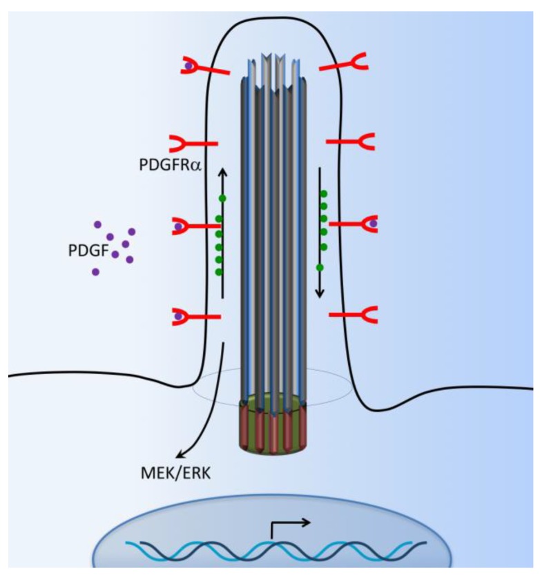
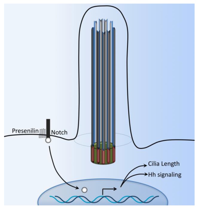
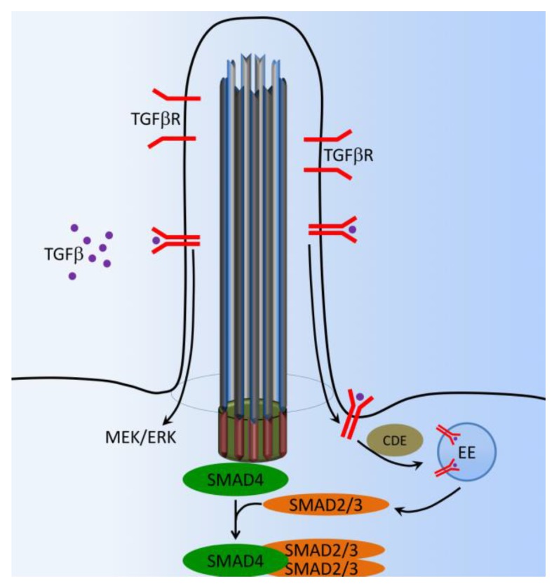
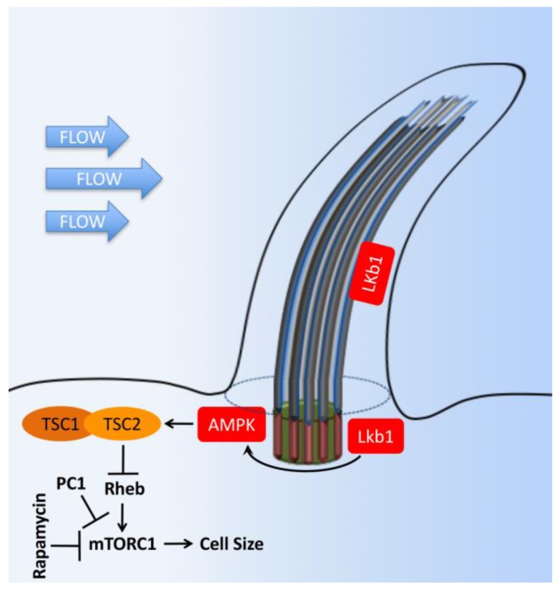
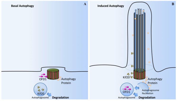
Primary cilia are hair-like organelles and play crucial roles in vertebrate development, organogenesis, health, and many genetic disorders. A primary cilium is a mechano-sensory organelle that responds to mechanical stimuli in the micro-environment. A cilium is also a chemosensor that senses chemical signals surrounding a cell. The overall function of a cilium is therefore to act as a communication hub to transfer extracellular signals into intracellular responses. Although intracellular calcium has been one of the most studied signaling messengers that transmit extracellular signals into the cells, calcium signaling by various ion channels remains a topic of interest in the field. This may be due to a broad spectrum of cilia functions that are dependent on or independent of utilizing calcium as a second messenger. We therefore revisit and discuss the calcium-dependent and calcium-independent ciliary signaling pathways of Hedgehog, Wnt, PDGFR, Notch, TGF-β, mTOR, OFD1 autophagy, and other GPCR-associated signaling. All of these signaling pathways play crucial roles in various cellular processes, such as in organ and embryonic development, cardiac functioning, planar cell polarity, transactivation, differentiation, the cell cycle, apoptosis, tissue homeostasis, and the immune response.

Keywords: calcium; cilioplasm; cytoplasm; sensory function; signaling.


Conflict of interest statement

The authors declare no conflict of interest.

Figures

Figure 1
Figure 1
Structure of a cilium. The cilium is a hair-like structure composed of the ciliary membrane, cilioplasm, axoneme, and basal body. This sensory organelle is membrane-bound and contains multiple microtubules running along its length. The ciliary membrane and axoneme make up the upper part of the cilium. The axoneme has nine peripheral microtubule doublets. The axoneme may have two central microtubules (9+2 vs. 9+0 axoneme). The 9+2 cilia usually have dynein arms and radial spokes that link the microtubule doublets and are motile, whereas most 9+0 cilia lack dynein arms, radial spoke, and central sheath and are nonmotile (primary cilia). Some 9+0 cilia only lack the central microtubule and are motile (nodal cilia). The axoneme is enclosed in the ciliary membrane, which is distinct from the cell membrane. Between the cilia and the cell membrane, there is a hypothetical transition membrane surrounding the transition fibers. The transition fibers at the junction of the basal body act as a filter for molecules that can pass into or out of the primary cilium.
Figure 2
Figure 2
Cilia-dependent calcium signaling. (A) The standard established model suggests that primary cilia respond to force through mechanosensation. In this original model, fluid flow bends the cilium, which triggers the opening of calcium-sensitive channel proteins and allows Ca2+ to enter the cilium. Intracellular signaling cascades are activated by the Ca2+ influx, leading to altered gene expression on the left side of the embryo, or promoting water transport in the kidneys. (B) In another model, ciliary bending in response to fluid force does not open Ca2+ channels. Instead, Ca2+ influx, observed in previous experiments, might have been due to diffusion from the cell body or caused by a damaged cilium membrane in response to extreme levels of shear force.
Figure 3
Figure 3
Cilia-dependent Hedgehog signaling. (A) The Hedgehog (Hh) pathway in vertebrates utilizes cilium as a signal transduction compartment. In the absence of the Hh ligand, the PTCH1 receptor localizes to the cilium and is thought to block the entry of SMO into cilia. The kinesin Kif7 (the COS2 homologue) localizes to the base of the cilium, where it may form a complex with Gli transcription factors and other pathway components. Kif7 at the cilium base prevents Gli proteins’ enrichment within the cilium and promotes processing of GliRs. (B) Upon Hh binding, SMO moves to the ciliary membrane and Kif7 translocates into the primary cilium, thereby promoting Gli accumulation at the ciliary tip. Kif7 and SMP in the cilia may also block the function of SUFU. Activated Gli (GliA) is transported out of the cilium by the dynein motor and intraflagellar transport particles (IFT). X and T symbols denote inhibition of gene transcription and protein function, respectively.
Figure 4
Figure 4
Cilia-dependent Wnt signaling. The cilia/basal body may function as a regulatory switch to control the canonical and non-canonical Wnt signaling pathways. (A) In the absence of fluid flow, canonical Wnt signaling predominates. Wnt ligand binds to the co-receptors frizzled, dishevelled (DSH) is recruited to frizzled, and glycogen synthase kinase-3 (GSK3) is inactivated. β-catenin (β-cat) translocates to the nucleus, where it acts as a transcriptional co-activator with members of the LEF and TCF family and induces transcription of Wnt target genes such as cMYC, AXIN2, or L1CAM. (B) In non-canonical Wnt signaling, the mechanosensation by fluid flow causes intracellular Ca2+ increase and an increase inversin expression. Inversin resides in multiple locations in the cell as well as at the base of the cilium. Inversin targets cytoplasmic DSH for anaphase-promoting complex/cyclosome (APC/C)-dependent ubiquitylation and degradation, making it unavailable for canonical Wnt signaling.
Figure 5
Figure 5
Cilia-dependent PDGFR signaling. The G-protein coupled receptors such as platelet-derived growth factor receptor (PDGFRα) are located at the cilia membrane. The PDGFR pathway is initiated with PDGF ligand binding to their receptors and inducing cellular responses through downstream signaling pathways such as the MEK/ERK cascade.
Figure 6
Figure 6
Cilia-dependent Notch signaling. A single-transmembrane Notch forms a receptor complex with the eight-transmembrane presenilin. Upon activation, presenilin mediates the intramembranal cleavage of notch containing the cdc10/Ankyrin repeats. The Notch intracellular domain is then released to the nucleus to activate various gene transcriptions.
Figure 7
Figure 7
Cilia-dependent TGF-β signaling. In the presence of TGF-β, activated TGF-β receptors (TGF-β1 and TGF-β2) translocate from the ciliary membrane to the ciliary pocket (CiPo) for clathrin-dependent endocytosis (CDE) and activation of SMAD transcription factors 2/3 (SMAD2/3) in early endosomes (EE) at the ciliary base. SMAD2/3 forms a trimeric complex with SMAD4 for nuclear translocation and activation of TGF-β target genes. Ciliary TGF-β signaling also involves the activation of ERK1/2.
Figure 8
Figure 8
Cilia-dependent mTOR signaling. Flow shear stress mediates the bending of primary cilia and inhibition of mTORC1. Tumor suppressor kinase (LKB1) and AMP-dependent protein kinase (AMPK) are found in the cilia and basal body. Upon cilium bending by fluid flow, LKB1 is activated and transported into the basal body, where AMPK is phosphorylated. Furthermore, AMPK phosphorylates TSC2 to activate the TSC1–TSC2 complex. In turn, TSC1–TSC2 stimulates the GTPase activity of Rheb, preventing it from activating mTORC1. Otherwise, AMPK could also directly antagonize mTORC1. Polycystin-1 (PC1) may also exert its inhibitory effect on the mTORC1 pathway through its known interaction with TSC2. The T bars denote inhibition of protein function.
Figure 9
Figure 9
Cilia-dependent OFD1/Autophagy signaling. (A) In basal autophagy, ciliogenesis is prevented by the sequestering of IFT20 in autophagosomes and its autophagic degradation. OFD1, a protein that constrains cilium formation at centriolar satellites, is not affected by basal autophagy and ciliogenesis is therefore impaired. (B) Autophagy induced by serum deprivation promotes ciliogenesis through inactivation of OFD1, while sparing IFT20. Autophagy proteins are also confined to this sensory organelle, probably contributing to cilium organization. At the basal body, signals from the cilium stimulate autophagosome nucleation and OFD1 degradation.

Similar articles

Cited by

References

    1. Gherman A., Davis E.E., Katsanis N. The ciliary proteome database: An integrated community resource for the genetic and functional dissection of cilia. Nat. Genet. 2006;38:961–962. doi: 10.1038/ng0906-961. - DOI - PubMed
    1. Ishikawa H., Thompson J., Yates J.R., 3rd, Marshall W.F. Proteomic analysis of mammalian primary cilia. Curr. Biol. 2012;22:414–419. doi: 10.1016/j.cub.2012.01.031. - DOI - PMC - PubMed
    1. Satir P., Christensen S.T. Overview of structure and function of mammalian cilia. Annu. Rev. Physiol. 2007;69:377–400. doi: 10.1146/annurev.physiol.69.040705.141236. - DOI - PubMed
    1. Lee L. Mechanisms of mammalian ciliary motility: Insights from primary ciliary dyskinesia genetics. Gene. 2011;473:57–66. doi: 10.1016/j.gene.2010.11.006. - DOI - PubMed
    1. Venkatesh D. Primary cilia. J. Oral Maxillofac. Pathol. 2017;21:8–10. doi: 10.4103/jomfp.JOMFP_48_17. - DOI - PMC - PubMed