Review
. 2018 Mar;15(3):139-150.
doi: 10.1038/nrcardio.2017.165. Epub 2017 Nov 16.

Next-generation pacemakers: from small devices to biological pacemakers

Affiliations
Review

Next-generation pacemakers: from small devices to biological pacemakers

Eugenio Cingolani et al. Nat Rev Cardiol. 2018 Mar.

Abstract

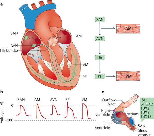
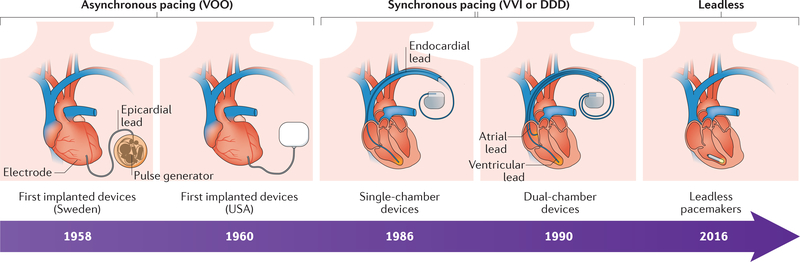
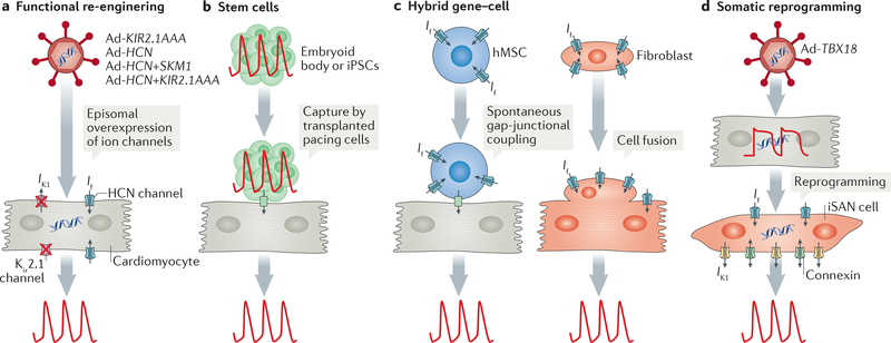
Electrogenesis in the heart begins in the sinoatrial node and proceeds down the conduction system to originate the heartbeat. Conduction system disorders lead to slow heart rates that are insufficient to support the circulation, necessitating implantation of electronic pacemakers. The typical electronic pacemaker consists of a subcutaneous generator and battery module attached to one or more endocardial leads. New leadless pacemakers can be implanted directly into the right ventricular apex, providing single-chamber pacing without a subcutaneous generator. Modern pacemakers are generally reliable, and their programmability provides options for different pacing modes tailored to specific clinical needs. Advances in device technology will probably include alternative energy sources and dual-chamber leadless pacing in the not-too-distant future. Although effective, current electronic devices have limitations related to lead or generator malfunction, lack of autonomic responsiveness, undesirable interactions with strong magnetic fields, and device-related infections. Biological pacemakers, generated by somatic gene transfer, cell fusion, or cell transplantation, provide an alternative to electronic devices. Somatic reprogramming strategies, which involve transfer of genes encoding transcription factors to transform working myocardium into a surrogate sinoatrial node, are furthest along in the translational pipeline. Even as electronic pacemakers become smaller and less invasive, biological pacemakers might expand the therapeutic armamentarium for conduction system disorders.


Conflict of interest statement

Competing interests statement

The authors declare no competing interests.

Figures

Figure 1 |
Figure 1 |. Cardiac conduction system.
a| Schematic representation of the anatomy and cellular electrophysiology of the cardiac conduction system. The cardiac impulse (arrows) originates in the sinoatrial node (SAN), travels across the atrial myocardium (AM), and moves through the atrioventricular node (AVN), the His bundle, and left and right bundle branches. The simultaneous activation of both bundle branches and their terminal Purkinje fibres (PF) provides antegrade activation of the ventricular myocardium (VM) in a synchronized fashion. b | Distinct action potential morphologies at different levels of the conduction system impose specialized electrical behaviours. c | Development of the SAN in mice. The SAN forms in the sinus venosus; during early embryonic development of the heart, transcription factors (green box) activate the SAN gene programme in the sinus venosus, while repressing the chamber myocyte gene programme. Chamber myocardium is depicted in purple, grey, and blue; non-chamber myocardium in dark pink; and non-myocardial tissue in light pink. ISL1, insulin gene enhancer protein ISL1; SHOX2, short stature homeobox protein 2; TBX, T-box transcription factor. Part c adapted from REF 9. http://creativecommons.org/licenses/by/3.0/.
Figure 2 |
Figure 2 |. Mechanisms of sinoatrial node automaticity.
A dual oscillator system consisting of a membrane clock and a calcium clock provides stable rhythmic depolarizations. The membrane clock relies on the pacemaker current (via hyperpolarization-activated cyclic nucleotide-gated (HCN) channels), L-type calcium channels, and T-type calcium channels for diastolic depolarization. The calcium clock works in synchrony with the membrane clock by releasing calcium from the sarcoplasmic reticulum (SR) through ryanodine receptors (RYRs) (step 1), thereby depolarizing the cell in diastole by activating the sodium-calcium exchanger (NCX) (step 2). The sarcoplasmic/endoplasmic reticulum calcium ATPase (SERCA), modulated by cardiac phospholamban (PLB), moves the calcium entering through the calcium channels in the plasma membrane back into the SR (step 3) to prime the calcium for the next cycle (step 4). Both the membrane clock and the calcium clock act in synchrony to create spontaneous depolarizations and are regulated by the autonomic nervous system through β-adrenergic and muscarinic stimulation, which modulates the activity of protein kinases (cAMP-dependent protein kinase (PKA) and calcium/calmodulin-dependent protein kinase type II (CAMKII)) that phosphorylate multiple proteins in the system. The calcium cycle is represented by the dashed grey arrows. ICaL, L-type calcium channel current; ICaT, T-type calcium channel current; If, funny current.
Figure 3 |
Figure 3 |. Timeline of the evolution of electronic pacemakers.
Early electronic pacemaker devices were big, had epicardial leads, and were implanted by open thoracotomy. Modern devices (single-chamber devices and dual-chamber devices) are much smaller, use endocardial leads, and can be implanted with minimally invasive techniques. First-generation devices provided only asynchronous (VOO) pacing, whereas modern devices are capable of synchronous and/or on-demand (VVI/DDD) pacing. Leadless pacemakers have a self-contained capsule that includes the battery, generator, and pacing electrodes, and are implanted percutaneously through the femoral vein.
Figure 4 |
Figure 4 |. Biological pacemaker approaches.
a | In a functional re-engineering approach, adenoviral (Ad) vectors are used to overexpress genes encoding ion channels (single channel or a combination of channels) in cardiomyocytes to create automaticity, for example, to increase the number of hyperpolarization-activated cyclic nucleotidegated (HCN) channels and to reduce the number of functional inward-rectifier potassium channels (Kir2.1) by overexpressing a dominant negative construct (KIR2.1AAA). b | With a stem cell approach, a cluster of beating cells derived from human embryonic stem cells (called an embryoid body) or induced pluripotent stem cells (iPSCs) are transplanted into a specific location in the heart to capture surrounding myocardium, thereby creating biological pacing. c | In a hybrid approach, cells (human mesenchymal stem cells (hMSCs) or fibroblasts) are used to deliver ion channel genes (for example, genes encoding components of HCN channels) to produce cardiac automaticity. Delivery by hMSCs requires gap-junctional coupling between the cardiomyocyte and the hMSC, whereas delivery by fibroblasts involves cell fusion. d | In somatic reprogramming, overexpression of the T-box transcription factor TBX18 by use of Ad vectors reprogrammes adult cardiac chamber cardiomyocytes into induced sinoatrial node (iSAN) cells, recapitulating the features of the SAN and therefore creating pacemaker activity. If, funny current; IK1, inward rectifier current.
Figure 5
Figure 5. Somatic reprogramming by TBX18.
a | A ventricular cardiomyocyte expressing T-box transcription factor TBX18 (TBX18-VM; middle) has a morphology resembling endogenous sinoatrial node (SAN) myocytes (left), in contrast to the rod shape of adult, non-reprogrammed green fluorescent protein (GFP)-expressing ventricular cardiomyocytes (GFP-VMs; right). b | Reprogrammed ventricular cardiomyocytes (TBX18-VM; top middle) have spontaneous oscillations, and the action potential (AP) morphology resembles that of endogenous SAN myocytes (top left). Non-reprogrammed ventricular cardiomyocytes (GFP-VM; top right) do not have spontaneous depolarizations and produce APs only in response to electrical stimulation. The bottom panels show AP recordings on a higher-resolution timescale. α-SA, α-smooth muscle actin; DAPI, 4’,6-diamidino-2-phenylindole; Em, membrane voltage. Figure from REF 11, Macmillan Publishers Limited.

Similar articles

Cited by

References

    1. Miranda JO, Ramalho C, Henriques-Coelho T & Areias JC Fetal programming as a predictor of adult health or disease: the need to reevaluate fetal heart function. Heart Fail. Rev. 22, 861–877 (2017). - PubMed
    1. Friedman D, Duncanson L, Glickstein J & Buyon J A review of congenital heart block. Images Paediatr. Cardiol. 5, 36–48 (2003). - PMC - PubMed
    1. Marban E Cardiac channelopathies. Nature 415, 213–218 (2002). - PubMed
    1. Bers DM Cardiac excitation-contraction coupling. Nature 415, 198–205 (2002). - PubMed
    1. Crick SJ et al. Innervation of the human cardiac conduction system. A quantitative immunohistochemical and histochemical study. Circulation 89, 1697–1708 (1994). - PubMed

Publication types

MeSH terms