. 2017 Nov 16;9(11):342.
doi: 10.3390/v9110342.

The Non-Homologous End Joining Protein PAXX Acts to Restrict HSV-1 Infection

Affiliations

The Non-Homologous End Joining Protein PAXX Acts to Restrict HSV-1 Infection

Ben J Trigg et al. Viruses. .

Abstract

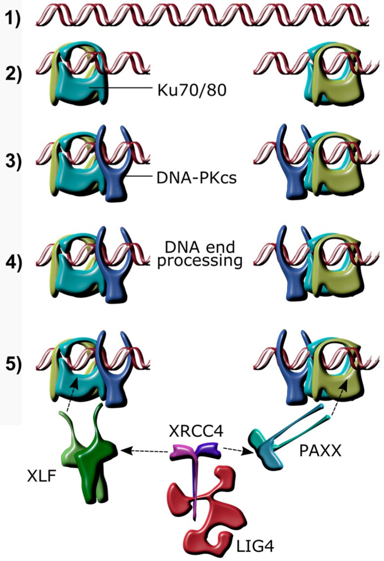
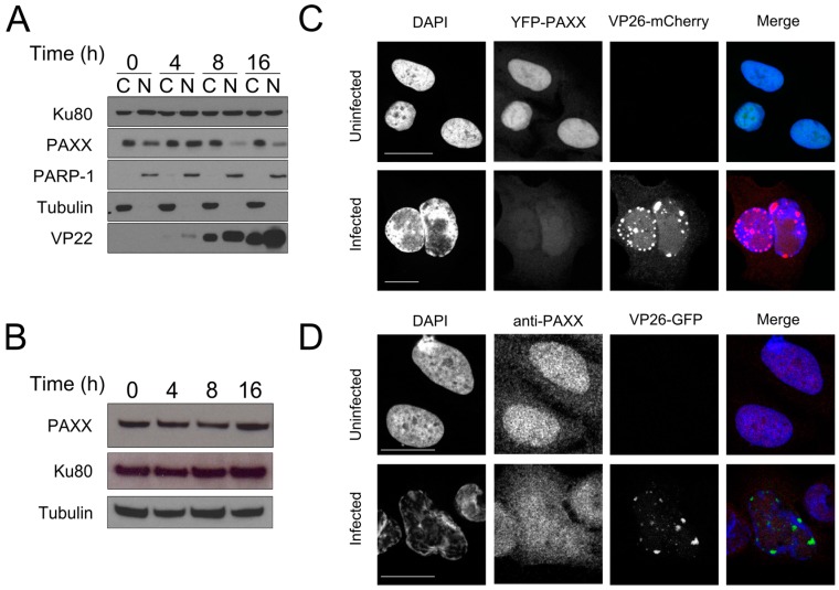
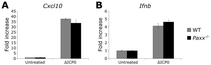
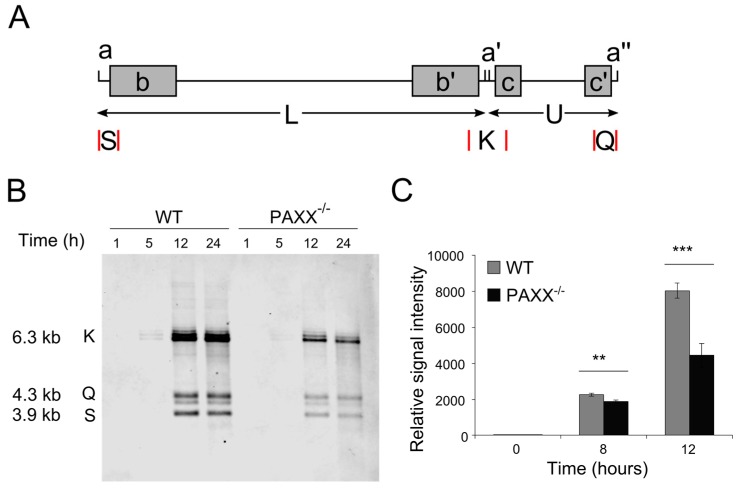
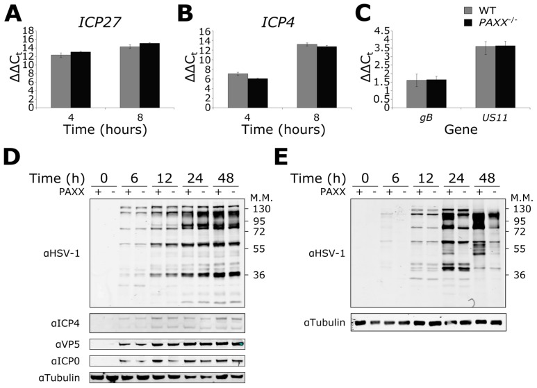
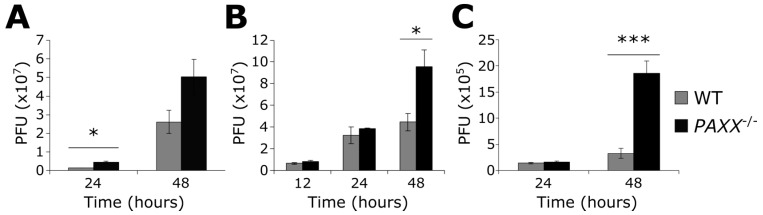
Herpes simplex virus 1 (HSV-1) has extensive interactions with the host DNA damage response (DDR) machinery that can be either detrimental or beneficial to the virus. Proteins in the homologous recombination pathway are known to be required for efficient replication of the viral genome, while different members of the classical non-homologous end-joining (c-NHEJ) pathway have opposing effects on HSV-1 infection. Here, we have investigated the role of the recently-discovered c-NHEJ component, PAXX (Paralogue of XRCC4 and XLF), which we found to be excluded from the nucleus during HSV-1 infection. We have established that cells lacking PAXX have an intact innate immune response to HSV-1 but show a defect in viral genome replication efficiency. Counterintuitively, PAXX-/- cells were able to produce greater numbers of infectious virions, indicating that PAXX acts to restrict HSV-1 infection in a manner that is different from other c-NHEJ factors.

Keywords: DDR; DNA damage response; HSV-1; PAXX; c-NHEJ; classical non-homologous end joining; herpes simplex virus 1.


Conflict of interest statement

The authors declare no conflict of interest.

Figures

Figure 1
Figure 1
Schematic overview of non-homologous end joining after a DNA double strand break. (1) Linear DNA; (2) A double strand break (DSB) caused by genotoxic damage is bound by the Ku70/80 heterodimer, which slides along the DNA to make room for subsequent complex formation; (3) DNA-dependent protein kinase, catalytic subunit (DNA-PKcs) binds to the Ku70/80 heterodimer; (4) Factors, including aprataxin, Aprataxin And PNKP Like Factor (APLF), Polynucleotide Kinase 3′-Phosphatase (PNKP), Artemis, Werner Syndrome RecQ Like Helicase (WRN), DNA polymerase lambda (Polλ), and DNA polymerase mu (Polμ) modify the ends of the DNA if necessary; (5) Paralogue of XRCC4 and XLF (PAXX) and/or XRCC4-like factor (XLF) bind Ku70/80 and recruit the XRCC4/Ligase 4 (LIG4) complex, which in turn ligates the DNA ends together.
Figure 2
Figure 2
PAXX distribution changes during HSV-1 infection. (A) U2OS cells were infected at a multiplicity of infection (MOI) of 10. Cells were harvested and the cytoplasmic (C) and nuclear (N) fractions separated and probed by immunoblotting for PAXX. Poly (ADP-Ribose)-Polymerase 1 (PARP-1) and tubulin controlled for fractionation, VP22 demonstrated successful infection, and Ku80 was used as a loading control; (B) U2OS cells were infected at MOI 10 and whole cell lysates immunoblotted with the indicated antibodies; (C) Yellow fluorescent protein (YFP)-PAXX was ectopically expressed in U2OS cells, and these were infected with HSV-1 VP26-mCherry at MOI 1. 16 h after infection slides were imaged by confocal microscopy; (D) U2OS cells were infected with ΔgE VP26-YFP HSV-1 at an MOI of 1 for 16 h and analysed by immunofluorescence. Scale bars in (C,D) denote 20 μm.
Figure 3
Figure 3
PAXX does not affect innate immune responses to HSV-1 ΔICP0 infection. WT or Paxx−/− Mouse embryo fibroblasts (MEFs) were infected with MOI 5 ΔICP0 for 5 hr, and qPCR was used to analyse the transcription of (A) Cxcl10; and (B) Ifnb. n = 3. Error bars denote mean +/− standard error of the mean (SEM).
Figure 4
Figure 4
PAXX does not affect the proportion of endless HSV-1 genomes, but promotes genome replication. (A) Linear structure of the HSV-1 genome, indicating repetitive elements (a, a′, a″) and the S, K, and Q fragments created by cleavage with BamHI (selected restriction sites are denoted by red lines); (B) WT and PAXX−/− cells were infected at MOI 4, and total cellular and viral DNA was isolated. The HSV-1 Q, K, and S fragments were visualised by southern blot following digestion with BamHI; (C) WT and PAXX−/− RPE-1 cells were infected at an MOI of 4, and genome copy numbers were quantified using TaqMan qPCR. n = 3. Statistical significance, calculated by Student’s t-test, is denoted by stars—** = p < 0.02, *** = p < 0.01. n = 3. Error bars denote mean +/− SEM.
Figure 5
Figure 5
Effects of PAXX on viral gene transcription and protein production. Wild type (WT) or PAXX−/− RPE-1 cells were infected at an MOI of 5, and RNA extracted. qPCR was used to quantify viral transcription of (A) ICP27; (B) ICP4; (C) gB and US11; (D) RPE-1 cells were infected at MOI 1 and harvested for protein at various times post infection. An anti-HSV-1 antibody was used, along with specific antibodies. (E) As in (D), but in MEFs. Error bars denote mean +/− SEM. n = 3.
Figure 6
Figure 6
PAXX restricts production of infectious HSV-1 virions. WT or PAXX−/− RPE-1 cells were infected with HSV-1 at MOI (A) 0.01, or (B) 4, and infectious virions were titrated onto vero cells to determine virus titre (plaque-forming units; PFU); (C) WT or Paxx−/− MEFs were infected with HSV-1 at an MOI of 0.01, and infectious virions were titrated onto vero cells. Statistical significance, calculated by Student’s t-test, is denoted by stars—* = p < 0.05, *** = p < 0.01. n = 3. Error bars denote mean +/− SEM.

References

    1. Lieber M.R., Ma Y., Pannicke U., Schwarz K. Mechanism and regulation of human non-homologous DNA end-joining. Nat. Rev. Mol. Cell Biol. 2003;4:712–720. doi: 10.1038/nrm1202. - DOI - PubMed
    1. Gottlieb T.M., Jackson S.P. The DNA-dependent protein kinase: Requirement for DNA ends and association with Ku antigen. Cell. 1993;72:131–142. doi: 10.1016/0092-8674(93)90057-W. - DOI - PubMed
    1. Yaneva M., Kowalewski T., Lieber M.R. Interaction of DNA-dependent protein kinase with DNA and with Ku: Biochemical and atomic-force microscopy studies. EMBO J. 1997;16 doi: 10.1093/emboj/16.16.5098. - DOI - PMC - PubMed
    1. Mimori T., Hardin J.A. Mechanism of interaction between Ku protein and DNA. J. Biol. Chem. 1986;261:10375–10379. - PubMed
    1. Buck D., Malivert L., de Chasseval R., Barraud A., Fondanèche M.C., Sanal O., Plebani A., Stéphan J.L., Hufnagel M., le Deist F., et al. Cernunnos, a novel nonhomologous end-joining factor, is mutated in human immunodeficiency with microcephaly. Cell. 2006;124:287–299. doi: 10.1016/j.cell.2005.12.030. - DOI - PubMed

LinkOut - more resources