. 2017 Nov 16;10(4):89.
doi: 10.3390/ph10040089.

Propagation of Fibrillar Structural Forms in Proteins Stopped by Naturally Occurring Short Polypeptide Chain Fragments

Affiliations

Propagation of Fibrillar Structural Forms in Proteins Stopped by Naturally Occurring Short Polypeptide Chain Fragments

Irena Roterman et al. Pharmaceuticals (Basel). .

Abstract

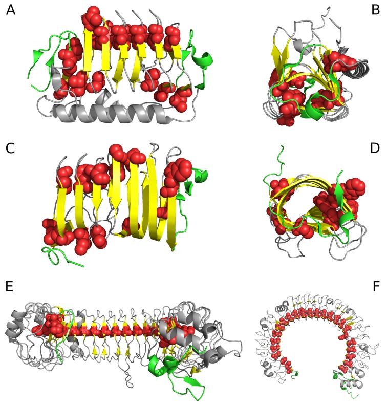
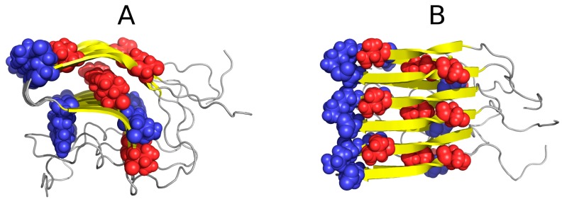
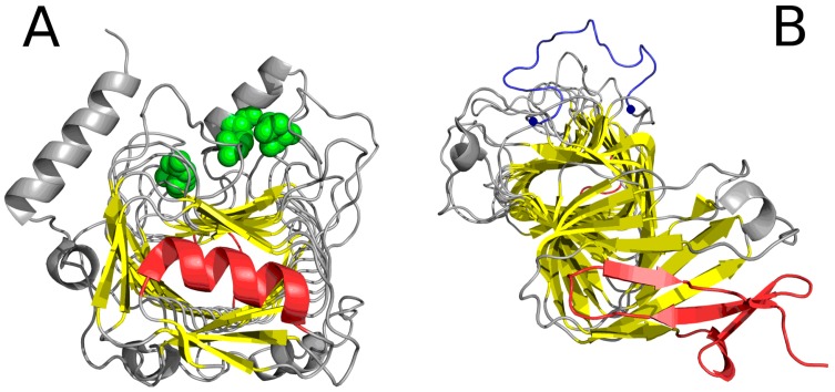
Amyloids characterized by unbounded growth of fibrillar structures cause many pathological processes. Such unbounded propagation is due to the presence of a propagating hydrophobicity field around the fibril's main axis, preventing its closure (unlike in globular proteins). Interestingly, similar fragments, commonly referred to as solenoids, are present in many naturally occurring proteins, where their propagation is arrested by suitably located "stopper" fragments. In this work, we analyze the distribution of hydrophobicity in solenoids and in their corresponding "stoppers" from the point of view of the fuzzy oil drop model (called FOD in this paper). This model characterizes the unique linear propagation of local hydrophobicity in the solenoid fragment and allows us to pinpoint "stopper" sequences, where local hydrophobicity quite closely resembles conditions encountered in globular proteins. Consequently, such fragments perform their function by mediating entropically advantageous contact with the water environment. We discuss examples of amyloid-like structures in solenoids, with particular attention to "stop" segments present in properly folded proteins found in living organisms.

Keywords: amyloid; fibrillation; hydrophobicity; solenoid.


Conflict of interest statement

The authors declare no conflict of interest.

Figures

Figure 1
Figure 1
Selected proteins viewed from two different angles. Left column: visualization of linear propagation of hydrophobic residues responsible for local maxima. Right column: visualization of “stop” fragments. (A,B): 3UYV; (C,D): 4YZA; (E,F): 2A0Z. In 2A0Z, the only residue directed toward the center is ILE 510. This protein is not included in our analysis due to its peculiar structural form which does not permit analysis based on the fuzzy oil drop model; however, it is visualized in the figure to show linear propagation of hydrophobic residues. Green fragments: stop fragments. Red space filling presentation: the positions of residues identified by fuzzy oil drop model as highly discordant versus the idealized distribution. This aims to show that they generate the linear propagation accordant to long axis of the solenoid. Gray fragments: additional chain fragments increasing the solubility of protein under consideration. However, this subject is not the object of analysis in this paper. Left column: solenoid axis—parallel to the paper sheet plane (A,C,E), right column—solenoid axis perpendicular to the paper sheet plane (B,D,F).
Figure 2
Figure 2
Example of an amyloid fibril (prion protein 2KJ3) in two projections, showing linear distribution of hydrophobicity (red: hydrophobic residues; blue: hydrophilic residues). Since no “stop” signal is present, the structure is susceptible to further linear aggregation. The regularity and of fibril without any polypeptide chain fragment disturbing the linear organization. (A,B)—different perspective: (A)—the fibril long axis—perpendiculat ro the paper sheet, (B)—the fibril axis parallel to the paper sheet.
Figure 3
Figure 3
Theoretical (blue), observed (red) and intrinsic (green) hydrophobicity distributions for successive β-sheets forming the lyase solenoid fragment (1DBG). Labels (A,B,C) follow the convention used to identify sheets in PDB. The blue line (T) shows the theoretical concentration of hydrophobicity in the central part, along with low hydrophobicity in the N- and C-terminal section, consistent with the 3D Gaussian. Proteins which exhibit such distribution are water-soluble. In contrast, the red line represents a distribution where no central peak can be observed and where hydrophobicity does not taper off in the terminal section—such as in the case of solenoids. Actual distribution (green) is closely aligned with the red profile, suggesting that the solenoid does not generate a central hydrophobic core and that it is moreover capable of complexing additional hydrophobic molecules in its terminal sections. This phenomenon is thought to be responsible for structural changes leading to formation of fibrillar forms rather than monocentric globules.
Figure 4
Figure 4
Hydrophobic (A) and hydrophilic (B) residues in 1DBG. VdW presentation has been applied in both images to highlight selected residues. Note the linear arrangement of hydrophobic and hydrophilic bands. The green helix at the bottom acts as a “stopper”.
Figure 5
Figure 5
“Stop” signals which accompany solenoids. (A) lyase (1DBG): red fragments: helix preventing further propagation of the fibrillar structure; green: residues engaged in enzymatic activity; yellow: β-sheet comprising the solenoid; (B) cell adhesion protein (1DAB): red fragments which constitute “stop” signals—an uncoiled loop (front) and a beta fold (red)—preventing propagation of fibrillar forms in either direction; blue: fragment (limited by two blue dots) believed to mediate interaction with epithelial cells [13] (GGXXP)5.
Figure 6
Figure 6
Examples of “stoppers” adjacent to the N- and C-terminal sections of the solenoid. Blue—theoretical distribution, red—observed one. (A) N-term 1L0S, (B) C-term 3UYV, (C) N-term 1DBG, (D) C-term 1L1I, (E) N-term 1EWW, (F) C-term 4YZA.
Figure 7
Figure 7
One-dimensional representation of fuzzy oil drop model parameters. The leftmost chart (A) presents the idealized Gaussian distribution—T—while the chart on the right (C) corresponds to the uniform distribution—R. Actual hydrophobicity distribution (expressed by the RD parameter) for the target protein is shown in the center (B) and marked on the axis with a pink dot (D). According to the fuzzy oil drop (FOD) model this protein contains a well-defined hydrophobic core. Vertical axes represent hydrophobicity (in arbitrary units), while horizontal axes represent distance (in multiplicities of σx). According to the three-sigma rule, the range between 0 + 3σ and 0 − 3σ covers more than 99% of the entire probability expressed by the Gaussian. The bottom axis shows the full range of the RD coefficient from 0 (perfect Gaussian) to 1 (uniform distribution with no concentration of hydrophobicity at any point in the protein body). In the presented example, RD = 0.318. RD > 0.5 is interpreted as a better match for the unified distribution than the theoretical Gaussian, whereas RD < 0.5 reveals the presence of a FOD-compliant monocentric hydrophobic core encapsulated in a hydrophilic shell.

Similar articles

Cited by

References

    1. Prusiner S.B. Novel proteinaceous infectious particles cause scrapie. Science. 1982;216:136–144. doi: 10.1126/science.6801762. - DOI - PubMed
    1. Prusiner S.B. Detecting mad cow disease. Sci. Am. 2004;291:86–93. doi: 10.1038/scientificamerican0704-86. - DOI - PubMed
    1. Stöhr J., Watts J.C., Mensinger Z.L., Oehler A., Grillo S.K., DeArmond S.J., Prusiner S.B., Giles K. Purified and synthetic Alzheimer’s amyloid beta (Aβ) prions. Proc. Natl. Acad. Sci. USA. 2012;109:11025–11030. doi: 10.1073/pnas.1206555109. - DOI - PMC - PubMed
    1. Taubes G. Misfolding the way to disease. Science. 1996;271:1493–1495. doi: 10.1126/science.271.5255.1493. - DOI - PubMed
    1. Purkey H.E., Dorrell M.I., Kelly J.W. Evaluating the binding selectivity of transthyretin amyloid fibril inhibitors in blood plasma. Proc. Natl. Acad. Sci. USA. 2001;98:5566–5571. doi: 10.1073/pnas.091431798. - DOI - PMC - PubMed

LinkOut - more resources