Case Reports
. 2017 Nov 16;551(7680):327-332.
doi: 10.1038/nature24487. Epub 2017 Nov 8.

Regeneration of the entire human epidermis using transgenic stem cells

Affiliations
Case Reports

Regeneration of the entire human epidermis using transgenic stem cells

Tobias Hirsch et al. Nature. .

Abstract

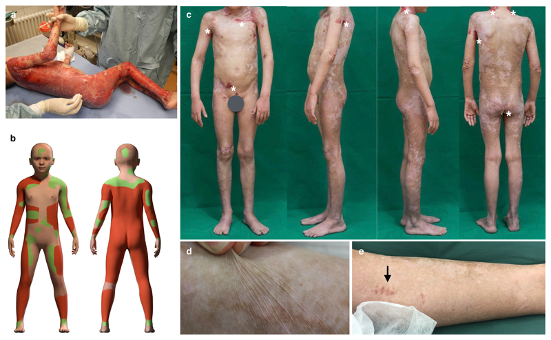
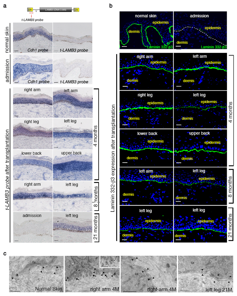
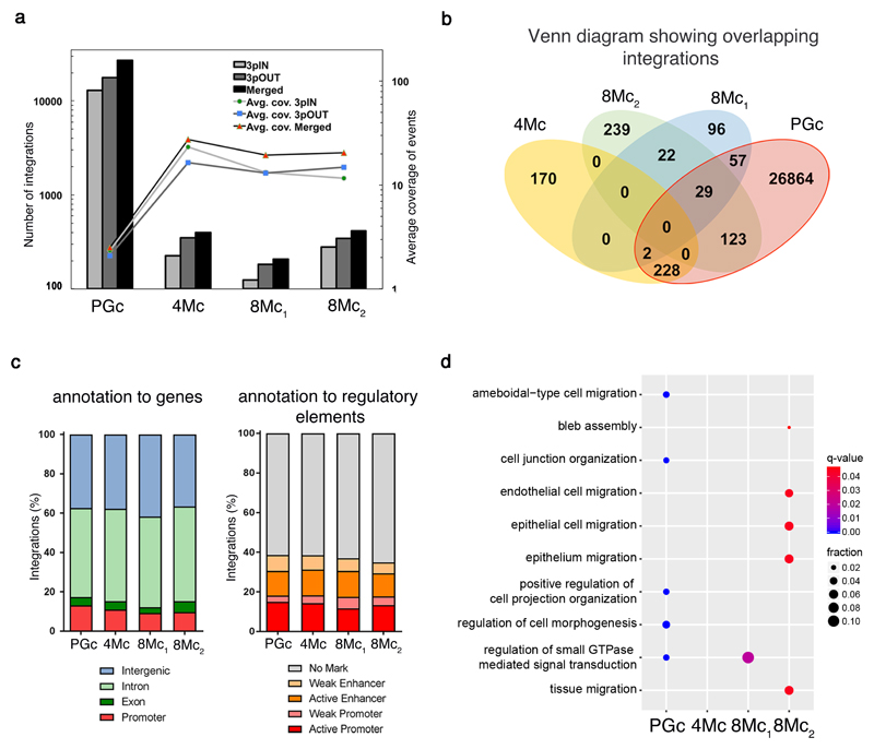
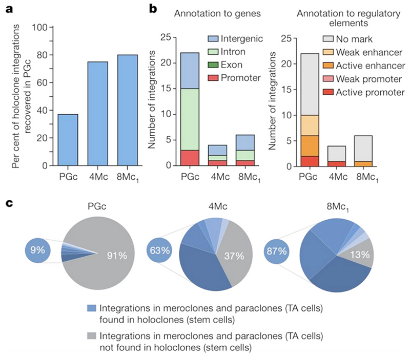
Junctional epidermolysis bullosa (JEB) is a severe and often lethal genetic disease caused by mutations in genes encoding the basement membrane component laminin-332. Surviving patients with JEB develop chronic wounds to the skin and mucosa, which impair their quality of life and lead to skin cancer. Here we show that autologous transgenic keratinocyte cultures regenerated an entire, fully functional epidermis on a seven-year-old child suffering from a devastating, life-threatening form of JEB. The proviral integration pattern was maintained in vivo and epidermal renewal did not cause any clonal selection. Clonal tracing showed that the human epidermis is sustained not by equipotent progenitors, but by a limited number of long-lived stem cells, detected as holoclones, that can extensively self-renew in vitro and in vivo and produce progenitors that replenish terminally differentiated keratinocytes. This study provides a blueprint that can be applied to other stem cell-mediated combined ex vivo cell and gene therapies.


Conflict of interest statement

Competing financial interests G.P. and M.D.L. are co-founders and member of the Board of Directors of Holostem Terapie Avanzate (HTA), s.r.l, Modena, Italy; Chiesi Farmaceutici S.p.A. (a co-founder of HTA), holds an Orphan Drug Medicinal Product designation (EU/3/15/1465) for the transgenic cultures used in this paper.

Figures

Figure 1
Figure 1. Regeneration of the transgenic epidermis.
a, Clinical picture of the patient showing massive epidermal loss. b, Schematic representation of the clinical picture. The denuded skin is indicated in red, while blistering areas are indicated in green. Flesh-colored areas indicate currently non-blistering skin. Transgenic grafts were applied on both red and green areas. c, Restoration of patient’s entire epidermis, with the exception of very few areas on the right thigh, buttocks, upper shoulders/neck and left axilla (asterisks, altogether ≤2% of TBSA). d, Normal skin functionality and elasticity. e, Absence of blister formation at sites where some of post-graft biopsies were taken (arrow).
Figure 2
Figure 2. Restoration of a normal epidermal-dermal junction.
Skin sections were prepared from normal skin, patient’ affected (admission) and transgenic skin at 4, 8 and 21 months follow-up. a, In situ hybridization was performed using a transgene-specific probe (t-LAMB3) on 10-μm-thick sections. E-cadherin-specific probe (Cdh1) was used as a control. Scale bars, 40 μm. b, Immunofluorescence of laminin 332-β3 was performed with 6F12 moAbs on 7-μm-thick sections. DAPI (blue) marks nuclei. Dotted line marks the epidermal-dermal junction. Scale bars, 20 μm. c, Electron-microscopy was performed on 70-nm-thick skin sections. A regular basement membrane (arrows) and normal hemidesmosomes (arrowheads, higher magnification in the inset) are evident in patient’ transgenic skin. Scale bars, 1 μm.
Figure 3
Figure 3. Integration profile of transgenic epidermis.
a, Integrations were identified in libraries obtained using two LTR-primers (3pIN, light grey bars; 3pOUT, dark grey bars; Supplementary Table 1) and in the merged set (black bars). Lines (secondary axis) depict the average integration coverage, calculated after removal of PCR duplicates. b, Venn diagram of the number of shared integrations across samples. c, percentage of integrations mapped to: promoters, exons, introns, and intergenic regions (left); epigenetically defined active and weak promoters and enhancers, or genomic regions with no histone marks (right); (p-value>0.05; Pearson's Chi-squared test). d, Dot plot of the top 5 enriched GO Biological Process terms for each sample. Dot colour indicates statistical significance of the enrichment (q-value); dot size represents the fraction of genes annotated to each term.
Figure 4
Figure 4. Integration profile of stem and TA cells.
a, Percentage of holoclone integrations recovered in the PGc bulk population. b, Holoclone integrations mapped to: promoters, exons, and introns, and intergenic regions (left); epigenetically defined active and weak promoters and enhancers, or genomic regions with no histone marks (right). c, The PGc pie chart (grey segment) shows that 91% of mero/paraclones did not contain the same integrations detected in the corresponding holoclones (each indicated by different blue segments). The 4Mc and 8Mc1 pie charts (grey segments) show that such percentage decreased to 37% and 17%, respectively.

Comment in

References

    1. Fine JD, et al. Inherited epidermolysis bullosa: updated recommendations on diagnosis and classification. J Am Acad Dermatol. 2014;70:1103–1126. doi: 10.1016/j.jaad.2014.01.903. - DOI - PubMed
    1. Fine JD, Johnson LB, Weiner M, Suchindran C. Cause-specific risks of childhood death in inherited epidermolysis bullosa. J Pediatr. 2008;152:276–280. doi: 10.1016/j.jpeds.2007.06.039. - DOI - PubMed
    1. Barrandon Y, Green H. Three clonal types of keratinocyte with different capacities for multiplication. Proceedings of the National Academy of Sciences of the United States of America. 1987;84:2302–2306. - PMC - PubMed
    1. Pellegrini G, et al. Location and clonal analysis of stem cells and their differentiated progeny in the human ocular surface. J Cell Biol. 1999;145:769–782. - PMC - PubMed
    1. Gallico GG, 3rd, O'Connor NE, Compton CC, Kehinde O, Green H. Permanent coverage of large burn wounds with autologous cultured human epithelium. The New England journal of medicine. 1984;311:448–451. doi: 10.1056/NEJM198408163110706. - DOI - PubMed

Publication types

MeSH terms

Substances