Review
. 2017 Nov 16;18(12):1288-1298.
doi: 10.1038/ni.3858.

Novel mechanisms and functions of complement

Affiliations
Review

Novel mechanisms and functions of complement

George Hajishengallis et al. Nat Immunol. .

Abstract

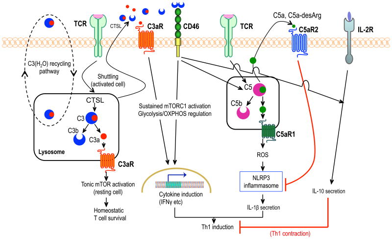
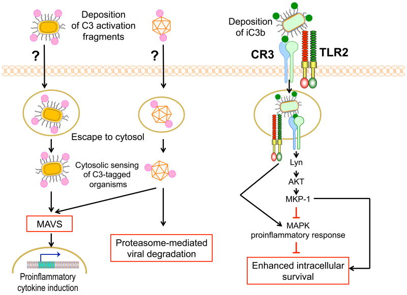
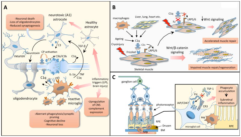
Progress at the beginning of the 21st century transformed the perception of complement from that of a blood-based antimicrobial system to that of a global regulator of immunity and tissue homeostasis. More recent years have witnessed remarkable advances in structure-function insights and understanding of the mechanisms and locations of complement activation, which have added new layers of complexity to the biology of complement. This complexity is readily reflected by the multifaceted and contextual involvement of complement-driven networks in a wide range of inflammatory and neurodegenerative disorders and cancer. This Review provides an updated view of new and previously unanticipated functions of complement and how these affect immunity and disease pathogenesis.


Figures

Figure 1
Figure 1. Complement-mediated intracellular and autocrine regulation of CD4+ T cell activation
Under steady-state conditions, C3(H2O) is recycling between the extracellular and intracellular space. The intracellular stores of C3(H2O) constitute a source of intracellular C3a. In resting CD4+ T cells, tonic intracellular C3a generation by cathepsin L (CTSL) cleavage activates the C3a receptor (C3aR) on lysosomes, leading to low-level mTOR activation for homeostatic T-cell survival. TCR activation with CD28 co-stimulation (not shown) causes translocation of this intracellular C3 activation system to the cell surface, where C3aR and CD46 are triggered, respectively, by C3a and C3b. The ensuing C3aR- and CD46-mediated signaling events stimulate sustained mTOR complex 1 (mTORC1) activation and reprogram glycolysis and oxidative phosphorylation in ways that support TH1 (IFN-γ) responses. In activated CD4+ T cells, CD46 costimulation triggers intracellular cleavage of C5 into C5a which induces C5aR1-dependent production of reactive oxygen species, in turn activating NLRP3 inflammasome-driven IL-1β secretion that sustains TH1 induction. IL-2R signaling together with autocrine CD46 activation induces IL-10 production, initiating transition to a TH1 contraction phase. This self-regulative activity is re-inforced by cell-surface C5aR2 signaling induced by increased levels of secreted C5a (or C5a-desArg) resulting from TH1 expansion; C5aR2 downregulates C5aR1-driven NLRP3 inflammasome activation.
Figure 2
Figure 2. Differential intracellular fate of microbes opsonized by complement in the extracellular space
(Left) Cytosolic sensing of C3-tagged microbes activates innate immunity. Certain intracellular pathogens can escape from phagosomes to replicate in the cytosol. However, nonenveloped viruses and bacteria, which were opsonized with C3 cleavage fragments in the extracellular space, can be sensed in the cytosol in a C3-dependent manner and trigger mitochondrial anti-viral signaling (MAVS) that leads to activation of innate immune responses. Cytosolic sensing of C3-opsonized viruses can additionally lead to proteasome-mediated viral degradation. The nature of the opsonizing C3 cleavage products and the uptake receptor(s) involved in this dual protective mechanism are uncertain. (Right) Immune evasion by intracellular pathogens dependent upon CR3 uptake. Several bacterial pathogens exploit CR3-mediated internalization into macrophages for enhanced intracellular persistence. For instance, CR3-mediated entry of F. tularensis induces CR3 signaling that activates Lyn kinase and AKT signaling, in turn upregulating MAP kinase phosphatase-1 (MKP-1) and thus attenuating MAP kinase-dependent pro-inflammatory responses downstream of TLR2.
Figure 3
Figure 3. Complement components implicated as drivers of chronic, non-resolving inflammation in neurodegenerative, aging-related and ocular pathologies
(a) An aberrantly reactivated complement-microglial axis promotes neurodegenerative phenotypes in the CNS. A hallmark of several neurological disorders is the complement-driven polarization of microglia and astrocytes towards proinflammatory phenotypes (A1 astrocytes) which impair synaptogenesis and compromise neuronal survival, as depicted herein and detailed in the text. (b) C1q promotes aging-related inflammation and tissue injury. C1q triggers the canonical Wnt pathway, independently of complement activation, by binding to the Wnt receptor Frizzled (Fz) and promoting the C1s-dependent cleavage of the Wnt coreceptor, LDL receptor-related protein 6 (LRP6). C1q-dependent enhancement of Wnt signaling accelerates fibrotic changes and impairs tissue regenerative responses (e.g., muscle repair). (c) Factor H promotes subretinal inflammation in age-related macular degeneration (AMD). Under steady-state conditions, phagocytic cells are constantly eliminated in the subretinal space through a homeostatic mechanism involving the interaction of TSP-1 with integrin-associated CD47 on subretinal phagocytes. The AMD-associated FH variant (H402) potently inhibits the CD47-driven elimination of phagocytes, presumably by binding more effectively to CR3 than the non-risk isoform, thereby fueling chronic subretinal inflammation. Abbreviations: RPE, retinal pigmented epithelium; BM, Bruch's membrane; Mφ, macrophage; TSP-1, thrombospondin-1; IAP, integrin-associated protein.
Figure 4
Figure 4. Role of complement in cancer
A complex interplay between complement effector proteins, tumor cells, and innate and adaptive immune cells in the tumor microenvironment determines tumor progression. Imbalanced complement activation in the tumor microenvironment triggers the release of proinflammatory cytokines by both tumor cells and tumor-infiltrating immune cells, such as macrophages, dendritic cells, and neutrophils, as well as immunosuppressive cytokines and reactive oxygen and nitrogen species by myeloid-derived suppressor cells (MDSCs). Local inflammation suppresses activation of effector T cells and creates a favorable environment for tumor growth. MDSCs also exert potent immunosuppressive effects that inhibit anti-tumor CD8+ T cell responses. Complement activation products, particularly C5a, promotes angiogenesis, thus facilitating tumor cell migration and adjacent tissue invasion and metastasis. Complement activation also triggers the accumulation of pro-tumorigenic neutrophils within solid tumors potentiating their procoagulant responses by releasing neutrophil extracellular traps (NETs).
Figure 5
Figure 5. Molecular mechanisms of C3-mediated complement activation, amplification, and effector generation
After initial activation of C3, via the classical (CP) or lectin pathways (LP), or other triggers, C3b is deposited on the cell surface and interacts with factor B (FB). The resulting proconvertase (C3bB) is subsequently activated by factor D (FD) to generate the alternative pathway C3 convertase, C3bBb. In the absence of regulators, an amplification loop driven by C3b deposition and convertase formation feeds into the terminal effector pathway by activating C5 and generating the C5a anaphylatoxin and the membrane attack complex (MAC). On host cells, regulators of complement activation (RCA) destabilize the C3 convertase and enable the binding of factor I (FI) to C3b, leading to its degradation to iC3b and C3dg. Opsonins resulting from this breakdown pathway interact with different complement receptors (CR) and exert distinct effector functions as indicated. The following Protein Data Bank (www.rcsb.org) structures were used: 2A73, 2I07, 4HW5, 1C3D, 2WIN, 2XWJ, 2XWB, 5O32, 2OK5, 1DSU, 2XRC, 3CU7, 1KJS, 4A5W. A hypothetical iC3b model was prepared using PDB 1C2D and 2A74. FH1-4 from PDB 2WII was used to visualized RCAs. The MAC was derived from the Electron Microscopy Data Bank (http://www.ebi.ac.uk/pdbe/emdb; EMD-3135).

Similar articles

Cited by

References

    1. Ricklin D, Hajishengallis G, Yang K, Lambris JD. Complement: a key system for immune surveillance and homeostasis. Nat Immunol. 2010;11:785–797. - PMC - PubMed
    1. Arbore G, Kemper C, Kolev M. Intracellular complement - the complosome - in immune cell regulation. Mol Immunol. 2017;89:2–9. - PMC - PubMed
    1. Ricklin D, Reis ES, Lambris JD. Complement in disease: a defence system turning offensive. Nat Rev Nephrol. 2016;12:383–401. - PMC - PubMed
    1. Elmlund D, Le SN, Elmlund H. High-resolution cryo-EM: the nuts and bolts. Curr Opin Struct Biol. 2017;46:1–6. - PubMed
    1. Ricklin D, Reis ES, Mastellos DC, Gros P, Lambris JD. Complement component C3 - The “Swiss Army Knife” of innate immunity and host defense. Immunol Rev. 2016;274:33–58. - PMC - PubMed

MeSH terms

Substances