

Pseudoalteromonas piratica strain OCN003 is a coral pathogen that causes a switch from chronic to acute Montipora white syndrome in Montipora capitata
- PMID: 29145488
- PMCID: PMC5690655
- DOI: 10.1371/journal.pone.0188319
Pseudoalteromonas piratica strain OCN003 is a coral pathogen that causes a switch from chronic to acute Montipora white syndrome in Montipora capitata
Abstract
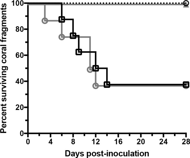
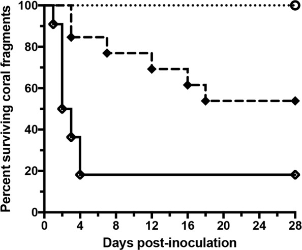
Reports of mass coral mortality from disease have increased over the last two decades. Montipora white syndrome (MWS) is a tissue loss disease that has negatively impacted populations of the coral Montipora capitata in Kāne'ohe Bay, Hawai'i. Two types of MWS have been documented; a progressive disease termed chronic MWS (cMWS), that can be caused by Vibrio owensii strain OCN002, and a comparatively faster disease termed acute MWS (aMWS), that can be caused by Vibrio coralliilyticus strain OCN008. M. capitata colonies exhibiting cMWS can spontaneously switch to aMWS in the field. In this study, a novel Pseudoalteromonas species, P. piratica strain OCN003, fulfilled Koch's postulates of disease causation as another etiological agent of aMWS. Additionally, OCN003 induced a switch from cMWS to aMWS on M. capitata in laboratory infection trials. A comparison of OCN003 and Vibrio coralliilyticus strain OCN008, showed that OCN003 was more effective at inducing the cMWS to aMWS switch in M. capitata than OCN008. This study is the first to demonstrate that similar disease signs on one coral species (aMWS on M. capitata) can be caused by multiple pathogens, and describes the first Pseudoalteromonas species that infects coral.
Conflict of interest statement
Figures


Similar articles
-
Vibrio coralliilyticus strain OCN008 is an etiological agent of acute Montipora white syndrome.Appl Environ Microbiol. 2014 Apr;80(7):2102-9. doi: 10.1128/AEM.03463-13. Epub 2014 Jan 24. Appl Environ Microbiol. 2014. PMID: 24463971 Free PMC article.
-
Emerging coral diseases in Kāne'ohe Bay, O'ahu, Hawai'i (USA): two major disease outbreaks of acute Montipora white syndrome.Dis Aquat Organ. 2016 May 26;119(3):189-98. doi: 10.3354/dao02996. Dis Aquat Organ. 2016. PMID: 27225202
-
Influence of salinity and sedimentation on Vibrio infection of the Hawaiian coral Montipora capitata.Dis Aquat Organ. 2018 Mar 22;128(1):63-71. doi: 10.3354/dao03213. Dis Aquat Organ. 2018. PMID: 29565254
-
Dynamics of acute Montipora white syndrome: bacterial communities of healthy and diseased M. capitata colonies during and after a disease outbreak.Microbiology (Reading). 2018 Oct;164(10):1240-1253. doi: 10.1099/mic.0.000699. Epub 2018 Jul 27. Microbiology (Reading). 2018. PMID: 30052176
-
Vibrio owensii induces the tissue loss disease Montipora white syndrome in the Hawaiian reef coral Montipora capitata.PLoS One. 2012;7(10):e46717. doi: 10.1371/journal.pone.0046717. Epub 2012 Oct 8. PLoS One. 2012. PMID: 23056419 Free PMC article.
Cited by
-
The Effect of Thermal Stress on the Physiology and Bacterial Communities of Two Key Mediterranean Gorgonians.Appl Environ Microbiol. 2022 Mar 22;88(6):e0234021. doi: 10.1128/aem.02340-21. Epub 2022 Mar 22. Appl Environ Microbiol. 2022. PMID: 35108095 Free PMC article.
-
Evolutionary Trajectory of the Replication Mode of Bacterial Replicons.mBio. 2021 Jan 26;12(1):e02745-20. doi: 10.1128/mBio.02745-20. mBio. 2021. PMID: 33500342 Free PMC article.
-
Case-control design identifies ecological drivers of endemic coral diseases.Sci Rep. 2020 Feb 18;10(1):2831. doi: 10.1038/s41598-020-59688-8. Sci Rep. 2020. PMID: 32071347 Free PMC article.
-
Disease Diagnostics and Potential Coinfections by Vibrio coralliilyticus During an Ongoing Coral Disease Outbreak in Florida.Front Microbiol. 2020 Oct 26;11:569354. doi: 10.3389/fmicb.2020.569354. eCollection 2020. Front Microbiol. 2020. PMID: 33193161 Free PMC article.
-
Genome sequence of Alteromonas macleodii strain OCN004 isolated from the extracellular mucus of an apparently healthy rice coral (Montipora capitata).Microbiol Resour Announc. 2024 Apr 11;13(4):e0007924. doi: 10.1128/mra.00079-24. Epub 2024 Feb 23. Microbiol Resour Announc. 2024. PMID: 38393331 Free PMC article.
References
-
- Antonius A. New observations on coral destruction in reefs. In University of Puerto Rico (Mayaguez); 1973.
-
- Woodley CM, Downs CA, Bruckner AW, Porter JW, Galloway SB. Diseases of Coral. John Wiley & Sons; 2016. 596 p.
-
- Harvell CD, Kim K, Burkholder JM, Colwell RR, Epstein PR, Grimes DJ, et al. Emerging Marine Diseases—Climate Links and Anthropogenic Factors. Science. 1999. September 3;285(5433):1505–10. - PubMed
-
- Jones RJ, Bowyer J, HoeghGuldberg O, Blackall LL. Dynamics of a temperature-related coral disease outbreak. Mar Ecol Prog Ser. 2004. November 1;281:63–77.
-
- Bourne DG. Microbiological assessment of a disease outbreak on corals from Magnetic Island (Great Barrier Reef, Australia). Coral Reefs. 2005. June 1;24(2):304–12.
MeSH terms
LinkOut - more resources
Full Text Sources
Other Literature Sources
Molecular Biology Databases
