Review
. 2018 Jan 1;159(1):32-45.
doi: 10.1210/en.2017-00882.

Immune System: An Emerging Player in Mediating Effects of Endocrine Disruptors on Metabolic Health

Affiliations
Review

Immune System: An Emerging Player in Mediating Effects of Endocrine Disruptors on Metabolic Health

Amita Bansal et al. Endocrinology. .

Abstract

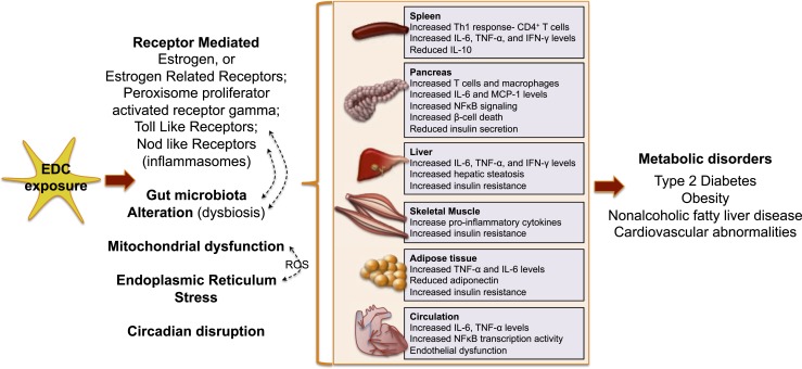
The incidence of metabolic disorders like type 2 diabetes and obesity continues to increase. In addition to the well-known contributors to these disorders, such as food intake and sedentary lifestyle, recent research in the exposure science discipline provides evidence that exposure to endocrine-disrupting chemicals like bisphenol A and phthalates via multiple routes (e.g., food, drink, skin contact) also contribute to the increased risk of metabolic disorders. Endocrine-disrupting chemicals (EDCs) can disrupt any aspect of hormone action. It is becoming increasingly clear that EDCs not only affect endocrine function but also adversely affect immune system function. In this review, we focus on human, animal, and in vitro studies that demonstrate EDC exposure induces dysfunction of the immune system, which, in turn, has detrimental effects on metabolic health. These findings highlight how the immune system is emerging as a novel player by which EDCs may mediate their effects on metabolic health. We also discuss studies highlighting mechanisms by which EDCs affect the immune system. Finally, we consider that a better understanding of the immunomodulatory roles of EDCs will provide clues to enhance metabolic function and contribute toward the long-term goal of reducing the burden of environmentally induced diabetes and obesity.


Figures

Figure 1.
Figure 1.
Role of immune system in mediating metabolic phenotype. The liver contains Kupffer cells (resident hepatic macrophages derived from yolk sac) and monocyte-derived recruited hepatic macrophages (recruited in liver from circulation), which become activated under metabolic distress and induce a proinflammatory phenotype by secreting a variety of inflammatory factors. This proinflammatory phenotype then contributes to hepatic steatosis and hepatic insulin resistance. Increased numbers of cytokines induce expression of genes involved in ceramide synthesis, an excess of which disrupts insulin signaling by inhibiting activation of Akt. In skeletal muscle, during metabolic distress, macrophages are recruited in myocytes. These accumulate within the intermyocyte adipocyte depots and in the surrounding vasculature, and secrete proinflammatory cytokines such as TNF, leading to decreased insulin signaling. In adipose tissue, activated adipocytes secrete adipokines and chemokines, which recruit monocytes (once recruited they become adipose-tissue macrophages) and proinflammatory invariant NK T cells, cytotoxic T cells (CD8+), and Th17 cells to adipocytes. The proinflammatory cells secrete proinflammatory TNF-α, IFN-γ, and IL-17, which polarize adipose-tissue macrophages to proinflammatory M1 macrophages. M1 macrophages with MHC II antigen presentation polarize naïve CD4+ cells to Th1 cells. Th1 cells, in turn, produce TNF-α and IFN-γ and further activate M1 macrophages, establishing a vicious cycle. The proinflammatory cytokines inhibit insulin signaling by direct serine phosphorylation of IRS1, thereby inducing insulin resistance. In the pancreas, in rodent models of type 2 diabetes and insulin resistance, macrophage infiltration into pancreatic islets is increased. Various signals within pancreatic islets activate macrophage NLRP3 inflammasomes, which subsequently cleave the proinflammatory IL-1 family of cytokines into their bioactive forms like IL-1β and IL-18, which bind to IL-1R1 on pancreatic islets, triggering inflammation-induced cell death. This eventually reduces insulin secretion. Furthermore, increased saturated fatty acid levels can induce lipotoxicity and β-cell apoptosis. In the intestine, in obesity and diabetes, the dysbiotic microbiota produces many metabolites, all of which can enter the circulation and negatively influence energy metabolism and insulin sensitivity. Additionally, altered metabolic state gastrointestinal leakiness can result in microbial products such as lipopolysaccharide gaining access to the circulation. Lipopolysaccharide in the bloodstream can contribute to insulin resistance by promoting tissue inflammation. In the circulatory system, minute cholesterol crystals in early atherosclerotic lesions activate the NLRP3 inflammasome in mmLDL-primed macrophages, promoting inflammatory cell infiltration and increased atherosclerosis. mmLDL, minimally modified low-density lipoprotein.
Figure 2.
Figure 2.
Inflammation-associated signaling pathways involved in insulin resistance. Proinflammatory signaling induces insulin resistance: Activation of TLR2 or TLR4 and/or TNFR initiates association of TAK1 and TAK1-binding protein 1, which, in turn, activates the NFκB IKK, JNK, and activator protein 1 pathways. Mitochondrial dysfunction leading to production of reactive oxygen species can also trigger IKK and JNK pathways, as can ER stress. Once triggered, IKK and JNK pathways activate serine kinase phosphorylation of IRS 1 and IRS2 and transcription of inflammatory genes. Together, these increase insulin resistance. Once assembled, the inflammasome activate caspase-1 subsequently cleaves the proinflammatory IL-1 family of cytokines into their bioactive forms, which further contribute to increased insulin resistance. Anti-inflammatory signaling reduces insulin resistance: Binding of omega-3 FAs activates GPR120, which initiates anti-inflammatory signaling by blocking TAK activation. Bonding of estrogen to estrogen receptor α (ERα) initiates anti-inflammatory response by blocking IKK–NFκB signaling. IL-10 binding to IL-10R also initiates anti-inflammatory signaling. The anti-inflammatory signals reduce insulin resistance. Akt, serine threonine kinase; ASC, apoptosis-associated speck-like protein containing a caspase recruitment domain; ER, endoplasmic reticulum; FA, fatty acid; GPR120, G-protein coupled receptor 120; IKK, inhibitor of κB kinase; IL-10R, IL-10 receptor; IRAK, interleukin 1 receptor associated kinase 1; Myd88, myeloid differentiation primary response gene 88; NLRP3, NOD-like receptor protein 3; PI3K, phosphoinositide 3-kinase; RIP, receptor interacting protein; SFA, saturated fatty acid; TAK1, transforming growth factor-β-activated kinase 1; TNFR, tumor necrosis factor receptor; TRADD, TNF receptor associated death domain; TRAF6, TNF receptor associated factor 6; TRIF, TIR domain containing adaptor protein inducing IFN-β; β-arr 2, β-arrestin 2.
Figure 3.
Figure 3.
Activation signals of inflammasomes. Signal 1 is provided by ligands of other pattern recognition receptors like TLR4, which, upon binding to exogenous pathogen-associated molecular patterns like LPSs, activates the downstream NFκB signaling pathway. NFκB translocates to the nucleus and induces expression of NLRP3 as well as other proinflammatory cytokines. Signal 2 includes organelle dysfunction (e.g., loss of plasma membrane integrity, lysosome rupture, mitochondrial dysfunction, ROS production, or autophagy induced by endoplasmic reticulum stress), invasion of microbial products (e.g., microbial protein, DNA), and perturbed homeostatic set points of cellular processes (e.g., aberrations in metabolites or influx of ions). Once assembled, the inflammasomes activate caspase-1, which subsequently cleaves the proinflammatory IL-1 family of cytokines into their bioactive forms, leading to pyroptosis. ASC, apoptosis-associated speck-like protein containing a caspase recruitment domain; ER, endoplasmic reticulum; NLRP3, NOD-like receptor protein 3; ROS, reactive oxygen species.
Figure 4.
Figure 4.
Possible routes of EDC action on the immune system contributing to metabolic disorders. By interacting with various receptors, altering the gut microbiome, inducing oxidative stress via mitochondrial dysfunction and/or endoplasmic reticulum stress, or via circadian disruption, EDCs trigger immune dysfunction in various tissues. Together, this may contribute toward a perturbed metabolic health. See Fig. 3 legend for expansion of abbreviation.

References

    1. Mathers CD, Loncar D. Projections of global mortality and burden of disease from 2002 to 2030. PLoS Med. 2006;3(11):e442. - PMC - PubMed
    1. American Diabetes Association Economic costs of diabetes in the U.S. in 2012 [published correction appears in Diabetes Care 2013;36(6):1797] Diabetes Care. 2013;36(4):1033–1046. - PMC - PubMed
    1. Finkelstein EA, Khavjou OA, Thompson H, Trogdon JG, Pan L, Sherry B, Dietz W. Obesity and severe obesity forecasts through 2030. Am J Prev Med. 2012;42(6):563–570. - PubMed
    1. Rowley WR, Bezold C, Arikan Y, Byrne E, Krohe S. Diabetes 2030: insights from yesterday, today, and future trends. Popul Health Manag. 2017;20(1):6–12. - PMC - PubMed
    1. Newbold RR, Padilla-Banks E, Jefferson WN, Heindel JJ. Effects of endocrine disruptors on obesity. Int J Androl. 2008;31(2):201–208. - PubMed

Publication types

MeSH terms

Substances