. 2017 Nov 16;17(1):769.
doi: 10.1186/s12885-017-3769-4.

Risk factors associated with the progression and metastases of hindgut neuroendocrine tumors: a retrospective study

Affiliations

Risk factors associated with the progression and metastases of hindgut neuroendocrine tumors: a retrospective study

Yoichiro Okubo et al. BMC Cancer. .

Abstract

Background: The worldwide incidence of neuroendocrine tumors (NETs) has increased remarkably, with the hindgut being the second most common site for such tumors. However, the mechanisms underlying progression and metastasis of hindgut NETs are unclear. A retrospective study was conducted to elucidate these mechanisms.

Methods: Clinicopathological data of cases of hindgut NET between April 1996 and September 2015 were analyzed, retrospectively. Patients with neuroendocrine carcinoma were excluded. Formalin-fixed paraffin-embedded tissues of hindgut NET cases were subjected to detailed morphometric and immunohistochemical analyses. Statistical analyses were performed using the non-parametric Mann-Whitney U test, Spearman's correlation coefficient, and chi-squared test. Multivariate logistic regression analysis was conducted as appropriate for the data set.

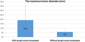
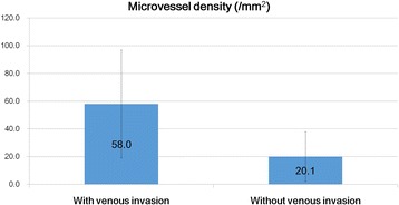
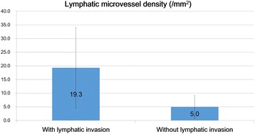
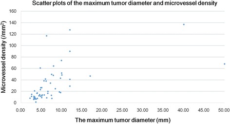
Results: Fifty-six hindgut NET cases were considered. Microvessel density and lymphatic microvessel density were identified as significant risk factors for venous and lymphatic invasion. There was a positive correlation between microvessel density and the maximum tumor diameter. Multivariate logistic regression analysis revealed that the maximum tumor diameter alone was an independent predictor of lymph node metastasis, whereas lymphovascular invasion and MVD was not the predictor of lymph node metastasis. There were no significant correlations between the Ki-67 labeling index and any of the parameters evaluated including age, sex, the maximum tumor diameter, venous invasion, lymphatic invasion, microvessel density, lymphatic microvessel density, and lymph node metastasis.

Conclusions: Angiogenic mechanisms may play important roles in the progression of hindgut NET. Otherwise, the maximum tumor diameter alone was an independent predictor of lymph node metastasis in hindgut NETs. Moreover, our study raises the question of whether the presence of lymphovascular invasion, in endoscopically obtained hindgut NET tissues, is an absolute indication for additional surgery or not.

Keywords: Angiogenesis; Hindgut; Lymphatic microvessel density; Lymphovascular invasion; Microvessel density; Neuroendocrine tumor.


Conflict of interest statement

Ethics approval and consent participate

This retrospective study was performed in accordance with the Declaration of Helsinki and approved by the Ethics Review Committee of Kanagawa Cancer Center, Kanagawa, Japan (Approval Number: 27–38). Furthermore, written informed consent was obtained at the time of clinical intervention for the future use of material for research in all cases.

Consent for publication

Not applicable.

Competing interests

The authors declare that they have no competing interests.

Publisher’s Note

Springer Nature remains neutral with regard to jurisdictional claims in published maps and institutional affiliations.

Figures

Fig. 1
Fig. 1
Representative images of histopathological findings in hindgut neuroendocrine tumors. a A photomicrograph showing a low-power field image of a hindgut neuroendocrine tumor (NET). The tumor cells are arranged in a trabecular pattern and show solid nests (Hematoxylin and eosin (HE) staining; original magnification, ×40; scale bar represents 1000 μm). b A photomicrograph showing a high-power field image of a hindgut NET. The tumor cells are uniform, arranged in rounded, solid nests, and have round-to-oval nuclei. Mild nuclear atypia can be seen (HE staining; original magnification, ×400; scale bar represents 100 μm)
Fig. 2
Fig. 2
Immunohistochemical reactivity for synaptophysin, chromogranin A, and Ki-67 in hindgut neuroendocrine tumors. Representative photomicrographs of immunohistochemical staining. a Tumor cells showed strong positive reactivity for synaptophysin (original magnification, ×100; scale bar represents 300 μm). b Tumor cells showed sporadic positive reactivity for chromogranin A (original magnification, ×100; scale bar represents 300 μm). c A few tumor cells showed positive reactivity for Ki-67 (original magnification, ×100; scale bar represents 300 μm)
Fig. 3
Fig. 3
Differences in the maximum tumor between tumors with and without lymph node metastasis. The maximum tumor diameter in patients with lymph node metastasis was significantly larger compared with that in those without lymph node invasion. The maximum tumor diameter was a significant risk factor for lymph node invasion in hindgut neuroendocrine tumors
Fig. 4
Fig. 4
Differences in microvessel density between tumors with and without venous invasion. The microvessel density (MVD) in tumors with venous invasion was significantly higher compared with that in tumors without venous invasion. MVD was a significant risk factor for venous invasion in hindgut neuroendocrine tumors (Mann–Whitney U test, P < 0.001; values are expressed as the mean ± standard deviation)
Fig. 5
Fig. 5
Differences in lymphatic microvessel density between tumors with and without lymphatic invasion. The lymphatic microvessel density in tumors with lymphatic invasion was significantly higher compared with that in tumors without lymphatic invasion. LMVD was a significant risk factor for lymphatic invasion in hindgut neuroendocrine tumors (Mann–Whitney U test, P < 0.001; values are expressed as the mean ± standard deviation)
Fig. 6
Fig. 6
Scatter plots of the hindgut neuroendocrine tumor between the maximum tumor diameter and microvessel density. A significant positive correlation was found between microvessel density and the maximum tumor diameter (r = 0.735, P < 0.001, Spearman correlation coefficient)

References

    1. Fraenkel M, Kim M, Faggiano A, de Herder WW, Valk GD, Knowledge N. Incidence of gastroenteropancreatic neuroendocrine tumours: a systematic review of the literature. Endocr Relat Cancer. 2014;21:R153–R163. doi: 10.1530/ERC-13-0125. - DOI - PubMed
    1. Hackeng WM, Hruban RH, Offerhaus GJ, Brosens LA. Surgical and molecular pathology of pancreatic neoplasms. Diagn Pathol. 2016;11:47. doi: 10.1186/s13000-016-0497-z. - DOI - PMC - PubMed
    1. Sevilla I, Segura A, Capdevila J, Lopez C, Garcia-Carbonero R, Grande E, et al. Management of controversial gastroenteropancreatic neuroendocrine tumour clinical situations with somatostatin analogues: results of a Delphi questionnaire panel from the NETPraxis program. BMC Cancer. 2016;16:858. doi: 10.1186/s12885-016-2901-1. - DOI - PMC - PubMed
    1. Okubo Y, Motohashi O, Nakayama N, Nishimura K, Kasajima R, Miyagi Y, et al. The clinicopathological significance of angiogenesis in hindgut neuroendocrine tumors obtained via an endoscopic procedure. Diagn Pathol. 2016;11:128. doi: 10.1186/s13000-016-0580-5. - DOI - PMC - PubMed
    1. Kim ST, Ha SY, Lee J, Hong SN, Chang DK, Kim YH, et al. The Clinicopathologic features and treatment of 607 hindgut neuroendocrine tumor (NET) patients at a single institution. Medicine (Baltimore) 2016;95:e3534. doi: 10.1097/MD.0000000000003534. - DOI - PMC - PubMed

LinkOut - more resources