. 2017 Nov 14;70(20):2504-2515.
doi: 10.1016/j.jacc.2017.09.036.

A Combination of Allogeneic Stem Cells Promotes Cardiac Regeneration

Affiliations

A Combination of Allogeneic Stem Cells Promotes Cardiac Regeneration

Makoto Natsumeda et al. J Am Coll Cardiol. .

Abstract

Background: The combination of autologous mesenchymal stem cells (MSCs) and cardiac stem cells (CSCs) synergistically reduces scar size and improves cardiac function in ischemic cardiomyopathy. Whereas allogeneic (allo-)MSCs are immunoevasive, the capacity of CSCs to similarly elude the immune system remains controversial, potentially limiting the success of allogeneic cell combination therapy (ACCT).

Objectives: This study sought to test the hypothesis that ACCT synergistically promotes cardiac regeneration without provoking immunologic reactions.

Methods: Göttingen swine with experimental ischemic cardiomyopathy were randomized to receive transendocardial injections of allo-MSCs + allo-CSCs (ACCT: 200 million MSCs/1 million CSCs, n = 7), 200 million allo-MSCs (n = 8), 1 million allo-CSCs (n = 4), or placebo (Plasma-Lyte A, n = 6). Swine were assessed by cardiac magnetic resonance imaging and pressure volume catheterization. Immune response was tested by histologic analyses.

Results: Both ACCT and allo-MSCs reduced scar size by -11.1 ± 4.8% (p = 0.012) and -9.5 ± 4.8% (p = 0.047), respectively. Only ACCT, but not MSCs or CSCs, prevented ongoing negative remodeling by offsetting increases in chamber volumes. Importantly, ACCT exerted the greatest effect on systolic function, improving the end-systolic pressure-volume relation (+0.98 ± 0.41 mm Hg/ml; p = 0.016). The ACCT group had more phospho-histone H3+ (a marker of mitosis) cardiomyocytes (p = 0.04), and noncardiomyocytes (p = 0.0002) than did the placebo group in some regions of the heart. Inflammatory sites in ACCT and MSC-treated swine contained immunotolerant CD3+/CD25+/FoxP3+ regulatory T cells (p < 0.0001). Histologic analysis showed absent to low-grade inflammatory infiltrates without cardiomyocyte necrosis.

Conclusions: ACCT demonstrates synergistic effects to enhance cardiac regeneration and left ventricular functional recovery in a swine model of chronic ischemic cardiomyopathy without adverse immunologic reaction. Clinical translation to humans is warranted.

Keywords: allogeneic; cardiac stem cell; ischemic cardiomyopathy; mesenchymal stem cell.


Figures

Figure 1
Figure 1. Suppressed negative remodeling of left ventricular volumes in response to ACCT treatment
From TESI at 3-months post-MI, to the end of the study (A) EDV increased in the placebo, CSC and MSC groups but remained unchanged in the ACCT group. (B) ESV increased in the placebo, CSC and MSC groups but remained unchanged in ACCT indicating that ACCT halts the progression of negative remodeling. *p<0.05, **p<0.01, ***p<0.001.
Figure 2
Figure 2. Cardiac function improved by combination stem cell therapy
Representative PV loops display the (A) decrease in ESPVR (in red), and increase in EDPVR (in blue) in placebo. In contrast, (B) ACCT produced the opposite effect. In response to TESI (C) the percent increase in ESPVR was significant only in ACCT, both within group and compared to placebo. (D) EDPVR and (E) EF did not improve significantly in any group. *p<0.05.
Figure 3
Figure 3. Myocardial perfusion of infarct zone improved with cell treatment
Perfusion MRI according to upslope analyses showed improvement of myocardial tissue perfusion in the infarct zone of ACCT compared to placebo-treated animals. ***p<0.001.
Figure 4
Figure 4. Increased mitosis of cardiomyocytes and non-cardiomyocytes in response to ACCT
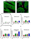
Confocal microscopy representative images of the mitosis specific marker, phospho-histone H3 (pHH3), in (A) cardiomyocytes and (B) non-cardiomyocytes. Based on the average number of pHH3+ cardiomyocytes in the (C) infarct zone (IZ), (D) border zone (BZ), and (E) remote zone (RZ), there was a two-fold increase in the RZ of ACCT, compared to placebo. pHH3+ non-cardiomyocytes in the (F) IZ, (G) BZ, and (H) RZ; there was a six-fold increase in the BZ of ACCT compared to placebo. *p<0.05, ****p<0.0001. DAPI, 4′, 6-diamidino-2-phenylindole.
Figure 5
Figure 5. Lymphocyte aggregates predominantly seen in injection sites of cell treated animals
Hematoxylin and eosin staining of extracted heart specimens demonstrate (A) lymphocyte aggregates in the BZ of an ACCT swine (scale bar, 200μm). (B) High magnification of delineated area shows lymphocytes confined to the interstitium without cardiomyocyte damage (scale bar, 50μm). Semi-quantitative analysis of 10 slides from each swine demonstrate lymphocyte aggregates were present in 20–25% of ACCT and MSC groups of (C) IZs (between group, p=0.98) and (D) BZs (between group, p=0.93). Lymphocyte aggregates was not seen in the (E) RZ. BZ, border zone; IZ, infarct zone, RZ, remote zone.
Figure 6
Figure 6. Tregs are expressed in all lymphocyte aggregates and granulomas
(A) Low magnification immunofluorescence confocal image demonstrates CD3+/CD25+/FoxP3+ Tregs in a lymphocyte aggregate from an ACCT-injected animal. (B–D) High magnification of the delineated area in panel A. Tregs (arrows) were confirmed in all lymphocyte aggregates and granulomas. Scale bars, 20μm. DAPI, 4′,6-diamidino-2-phenylindole; Tregs, regulatory T cells.
Central Illustration
Central Illustration. Scar mass and scar mass as percentage of left ventricular mass post-treatment
DE-MRI Short-axis images show the chronological change of scar size (scar=red) before (A,D,G,J) and 3-months after (B,E,H,K) treatment and, as seen in comparable gross histological sections (C,F,I,L). (M) Scar mass as a percentage of LV mass decreased significantly after TESI in ACCT and MSC. (N) Percent decrease in scar mass was greater in ACCT and MSC compared to placebo. *p<0.05, ***p<0.0001.

Comment in

References

    1. Quevedo HC, Hatzistergos KE, Oskouei BN, et al. Allogeneic mesenchymal stem cells restore cardiac function in chronic ischemic cardiomyopathy via trilineage differentiating capacity. Proc Natl Acad Sci U S A. 2009;106:14022–7. - PMC - PubMed
    1. Williams AR, Suncion VY, McCall F, et al. Durable scar size reduction due to allogeneic mesenchymal stem cell therapy regulates whole-chamber remodeling. J Am Heart Assoc. 2013;2:e000140. - PMC - PubMed
    1. Hatzistergos KE, Quevedo H, Oskouei BN, et al. Bone Marrow Mesenchymal Stem Cells Stimulate Cardiac Stem Cell Proliferation and Differentiation. Circulation Research. 2010;107:913. - PMC - PubMed
    1. McCall FC, Telukuntla KS, Karantalis V, et al. Myocardial infarction and intramyocardial injection models in swine. Nat Protoc. 2012;7:1479–96. - PMC - PubMed
    1. Heldman AW, DiFede DL, Fishman JE, et al. Transendocardial mesenchymal stem cells and mononuclear bone marrow cells for ischemic cardiomyopathy: the TAC-HFT randomized trial. Jama. 2014;311:62–73. - PMC - PubMed

LinkOut - more resources