Review
. 2018 Apr-May;223(4-5):383-396.
doi: 10.1016/j.imbio.2017.11.001. Epub 2017 Nov 12.

Macrophages: Their role, activation and polarization in pulmonary diseases

Affiliations
Review

Macrophages: Their role, activation and polarization in pulmonary diseases

Shweta Arora et al. Immunobiology. 2018 Apr-May.

Abstract

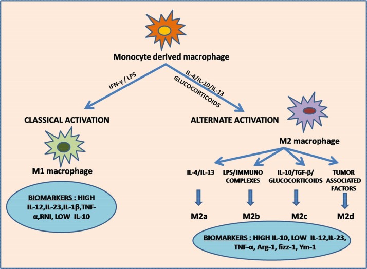
Macrophages, circulating in the blood or concatenated into different organs and tissues constitute the first barrier against any disease. They are foremost controllers of both innate and acquired immunity, healthy tissue homeostasis, vasculogenesis and congenital metabolism. Two hallmarks of macrophages are diversity and plasticity due to which they acquire a wobbling array of phenotypes. These phenotypes are appropriately synchronized responses to a variety of different stimuli from either the tissue microenvironment or - microbes or their products. Based on the phenotype, macrophages are classified into classically activated/(M1) and alternatively activated/(M2) which are further sub-categorized into M2a, M2b, M2c and M2d based upon gene expression profiles. Macrophage phenotype metamorphosis is the regulating factor in initiation, progression, and termination of numerous inflammatory diseases. Several transcriptional factors and other factors controlling gene expression such as miRNAs contribute to the transformation of macrophages at different points in different diseases. Understanding the mechanisms of macrophage polarization and modulation of their phenotypes to adjust to the micro environmental conditions might provide us a great prospective for designing novel therapeutic strategy. In view of the above, this review summarises the activation of macrophages, the factors intricated in activation along with benefaction of macrophage polarization in response to microbial infections, pulmonary toxicity, lung injury and other inflammatory diseases such as chronic obstructive pulmonary dysplasia (COPD), bronchopulmonary dysplasia (BPD), asthma and sepsis, along with the existing efforts to develop therapies targeting this facet of macrophage biology.

Keywords: Alternative activation; Asthma; BPD; COPD; Classical activation; Lung inflammation; M1/M2 macrophages.


Figures

Fig. 1
Fig. 1
Classical and Alternate pathway of Macrophage polarization. The figure depicts the pathways involved in macrophage polarization in response to signals received from micro-environment. Classical activation of M1 macrophages is induced by LPS/IFN-γ exposure. Activated M1 macrophages promote enhanced secretion of M1 chemokines, Th1 response elements, i-NOS (inducible nitric oxide synthase) dependent reactive nitrogen intermediates (RNI), high levels of IL-12, IL-23 IL-1β and TNF-α, and low levels of IL-10, which exert pro-inflammatory and cytotoxic effects. They are also involved in tumor suppression and immunostimulation. Alternately activated M2 macrophages are stimulated by IL-4, IL-10, IL-13 and glucocorticoids. IL-4 and IL-13 activates M2a subtype. Presence of immunocomplexes and LPS activates M2b subtype. M2c subtype is induced by IL-10, TGF-β and glucocorticoids. Presence of tumor associated factors triggers the activation of M2d subtype. Activated M2 macrophages enhance the secretion of IL-10 and reduces the secretion of IL-12 and IL-23 due to which they exert anti-inflammatory effects and roles in tissue repair and wound healing. M2d subtype is the prime constituent of TAMs (tumor associated macrophages) and hence promote tumor growth.
Fig. 2
Fig. 2
Transcription factors involved in macrophage polarization. The figure represents the role of transcription factors involved in M1/M2 polarization and their feedback control. M1 polarization: Binding of IFN-γ to its receptor activates STAT1, involved in transcription of IL-12, NOS-2 and MHC-II genes. Binding of LPS to TLR4 causes activation of IRF5, NF-κβ and AP1, all of which are involved in increased production of proinflammatory cytokines – IL-1, IL-6,IL-12,IL-23 and TNF. M2 polarization: Binding of free fatty acids to their receptors induces activation of PPARγ, involved in transcription of IL-10 and Arg-1 genes. IL-4/IL-13 triggers the activation of STAT6 and IRF4, responsible for transcription of Arg-1, IL-4α, Ym-1 and fizz-1 genes. TLR4/LPS binding activates CREB, which induces enhanced production of IL-10. M1/M2 polarization also exert feedback regulation mediated by STAT1-STAT6, IRF5-IRF4, NF-κβ-PPARγ, AP1-PPARγ and AP1-CREB. Blocked arrows represent feedback control.
Fig. 3
Fig. 3
Macrophage polarization – JAK/STAT pathway. Binding of IFNγ, LPS or IL-4/13 to their corresponding surface receptors triggers activation of JAKs (Janus Kinases) which induces activation of STATs (Signal transducer and activators of transcription) and transcription of M1and M2 genes. These genes are also transcribed by the differential activation of Akt1/2 via PI3K or PIP3. Binding of IFNγ to IFNγR1/2 activates JAK1/2, which in turn, activates STAT1/3. STAT1/3 induces activation of NF-κβ. Binding of LPS to TLR4 also activates NF-κβ via adapter proteins MyD88 (Myeloid differentiation primary response 88)/TRIF (TIR-domain containing adapter-inducing interferon-β). KLF4 inhibits the activity of NF-κβ. SOCS3 (suppressor of cytokine signalling 3) negatively regulates the cytokine signalling by binding to JAK2 kinase and inhibiting its activity. Binding of IL-4 to its receptor activates JAK1/2/3 kinases or PI3 kinases. Activated JAKs trigger activation of PPARγ/δ (Peroxisome proliferator-activated receptor gamma) via STAT6, which in turn, activates M2 genes. Akt1 activation promotes activation of M1 genes and Akt2 activation promotes activation of M2 genes.
Fig. 4
Fig. 4
Differential metabolism in macrophage polarization. This figure represents the metabolic differences in macrophage polarization. There is increased glycolytic activity induced by M1 macrophages which leads to higher production of lactate. Increase in NADPH is observed due to PPP (Pentose Phosphate Pathway) which contributes to the generation of ROS (reactive oxygen species) responsible for killing activity exerted by M1 cells. The cytotoxic activity of M1 cells is due to the production of NO (Nitric oxide) which is produced by catabolism of L-Arginine via i-NOS (Inducible Nitric oxide synthase) dependent pathway. M2 cells are involved in the catabolism of L-ornithine and polyamines mediated by Arg-1 expression. They also induce β oxidation, Krebs cycle and oxidative phosphorylation. Polyamines and L-Ornithine generated are involved in tissue repair and regeneration activity of M2 macrophages.
Fig. 5
Fig. 5
Macrophages in homeostasis. This figure portrays all the processes involved in cessation and repair of alveolar inflammation after acute inflammatory lung injury Homeostasis is a timely coordinated, active process in which alveolar macrophages are directly or indirectly involved. These processes include blockage of granulocyte (PMN) and monocyte influx from the circulation, phagocytosis of apoptotic polymorphonuclear neutrophils or parenchymal cells, initiation of angiogenesis, repair of the endo-and epithelial barrier by junctional sealing, clearance of alveolar edema, proliferation/differentiation of epithelial progenitor cells including type II alveolar epithelial cells (AEC), removal of fibrin and protein rich edema fluid. These processes are well synchronized and are critical for healthy tissue homeostasis.

References

    1. Agarwal R., et al. Etiology and outcomes of pulmonary and extrapulmonary acute lung injury/ARDS in a respiratory ICU in North India. Chest. 2006;130(3):724–729. - PubMed
    1. Alam K., et al. Glutathione-redox balance regulates c-rel-driven IL-12 production in macrophages: possible implications in antituberculosis immunotherapy. J. Immunol. 2010;184(6):2918–2929. - PubMed
    1. Amano H., et al. Essential contribution of monocyte chemoattractant protein-1/C-C chemokine ligand-2 to resolution and repair processes in acute bacterial pneumonia. J. Immunol. 2004;172(1):398–409. - PubMed
    1. Anderson C.F., Mosser D.M. A novel phenotype for an activated macrophage: the type 2 activated macrophage. J. Leukoc. Biol. 2002;72(1):101–106. - PubMed
    1. Arango Duque G., Descoteaux G.A. Macrophage cytokines: involvement in immunity and infectious diseases. Front. Immunol. 2014;5:491. - PMC - PubMed

Publication types