

Relative Contributions of the Midcarpal and Radiocarpal Joints to Dart-Thrower's Motion at the Wrist
- PMID: 29146510
- PMCID: PMC5837914
- DOI: 10.1016/j.jhsa.2017.10.017
Relative Contributions of the Midcarpal and Radiocarpal Joints to Dart-Thrower's Motion at the Wrist
Abstract
Purpose: To identify the relative contributions of the radiocarpal (RC) and midcarpal (MC) joints to dart-thrower's motion (DTM) of the wrist.
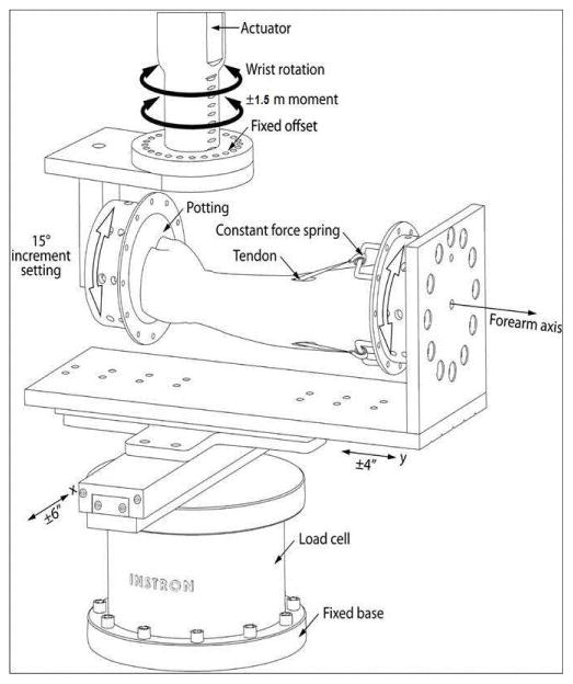
Methods: Six cadaveric upper extremities were fixed to a custom-designed loading jig allowing for pure moment-rotation analysis in 24 different directions of wrist motion. Each specimen was tested in 3 states: intact, simulated radiocarpal fusion (sRCF) and simulated pancarpal fusion (sPCF). Moments of ± 1.5 Nm were applied at each of 24 directions for each state and the resulting wrist rotation recorded. Data from each specimen were reduced to compute the range of motion (ROM) envelopes and the orientation of the ROM for the 3 different states.
Results: The ROM was significantly decreased in the sRCF and sPCF groups compared with the intact group in the directions of the pure extension, radial extension, ulnar flexion, and ulnar deviation. No significant difference in ROM was detected between the sRCF and sPCF groups in any direction. The ROM envelopes for the intact, sRCF, and sPCF groups were all oriented obliquely to the axis of pure wrist flexion-extension near a path of ulnar flexion-radial extension, consistent with prior reports on DTM.
Conclusions: Although both simulated fusion types decreased ROM compared with the intact wrist, the principal direction of wrist motion along the path of DTM was not significantly altered by simulated RCF or PCF.
Clinical relevance: These findings suggest that the RC and MC joints can each contribute to a similar mechanical axis of motion located along the path of DTM when the other joint has been eliminated via fusion. Surgical options such as partial wrist fusions may maintain the native wrist's mechanical axis if either the RC or the MC joint is preserved, despite significant reduction in overall ROM.
Keywords: Dart-thrower’s motion; midcarpal joint; radiocarpal fusion; wrist biomechanics.
Copyright © 2018 American Society for Surgery of the Hand. Published by Elsevier Inc. All rights reserved.
Figures


Similar articles
-
Simulated radioscapholunate fusion alters carpal kinematics while preserving dart-thrower's motion.J Hand Surg Am. 2008 Apr;33(4):503-10. doi: 10.1016/j.jhsa.2007.12.013. J Hand Surg Am. 2008. PMID: 18406953 Free PMC article.
-
In-vivo confirmation of the use of the dart thrower's motion during activities of daily living.J Hand Surg Eur Vol. 2014 May;39(4):373-8. doi: 10.1177/1753193412460149. Epub 2012 Sep 12. J Hand Surg Eur Vol. 2014. PMID: 22976876
-
Changes in Wrist Motion After Simulated Scapholunate Arthrodesis: A Cadaveric Study.J Hand Surg Am. 2016 Sep;41(9):e285-93. doi: 10.1016/j.jhsa.2016.07.056. J Hand Surg Am. 2016. PMID: 27570228
-
2007 IFSSH committee report of wrist biomechanics committee: biomechanics of the so-called dart-throwing motion of the wrist.J Hand Surg Am. 2007 Nov;32(9):1447-53. doi: 10.1016/j.jhsa.2007.08.014. J Hand Surg Am. 2007. PMID: 17996783 Review.
-
International Federation of Societies for Surgery of the Hand 2013 Committee's report on wrist dart-throwing motion.J Hand Surg Am. 2014 Jul;39(7):1433-9. doi: 10.1016/j.jhsa.2014.02.035. J Hand Surg Am. 2014. PMID: 24888529 Review.
Cited by
-
Dynamic MRI of the wrist in less than 20 seconds: normal midcarpal motion and reader reliability.Skeletal Radiol. 2020 Feb;49(2):241-248. doi: 10.1007/s00256-019-03266-1. Epub 2019 Jul 9. Skeletal Radiol. 2020. PMID: 31289900 Free PMC article.
-
In Vivo Measurement of Wrist Movements during the Dart-Throwing Motion Using Inertial Measurement Units.Sensors (Basel). 2021 Aug 20;21(16):5623. doi: 10.3390/s21165623. Sensors (Basel). 2021. PMID: 34451068 Free PMC article.
-
Proximal-distal shift of the center of rotation in a total wrist arthroplasty is more than twice of the healthy wrist.J Orthop Res. 2020 Jul;38(7):1575-1586. doi: 10.1002/jor.24717. Epub 2020 May 25. J Orthop Res. 2020. PMID: 32401391 Free PMC article.
-
Demystifying Palmar Midcarpal Instability.J Wrist Surg. 2021 Apr;10(2):94-101. doi: 10.1055/s-0040-1714688. Epub 2020 Aug 27. J Wrist Surg. 2021. PMID: 33815943 Free PMC article.
-
Biomechanical Comparison of Dart-Throw Motions after Partial Wrist Fusions.J Wrist Surg. 2021 Aug 5;11(1):69-75. doi: 10.1055/s-0041-1732412. eCollection 2022 Feb. J Wrist Surg. 2021. PMID: 35127267 Free PMC article.
References
-
- Palmer AK, Werner FW, Murphy D, Glisson R. Functional wrist motion: a biomechanical study. J Hand Surg Am. 1985;10(1):39–46. - PubMed
-
- Kauer JM. The mechanism of the carpal joint. Clin Orthop Relat Res. 1986;202:16–26. - PubMed
-
- Moritomo H, Murase T, Goto A, Oka K, Sugamoto K, Yoshikawa H. In vivo three-dimensional kinematics of the midcarpal joint of the wrist. J Bone Joint Surg Am. 2006;88(3):611–621. - PubMed
-
- Moritomo H, Apergis EP, Garcia-Elias M, Werner FW, Wolfe SW. International Federation of Societies for Surgery of the Hand 2013 Committee’s report on wrist dart-throwing motion. J Hand Surg Am. 2014;39(7):1433–1439. - PubMed
Publication types
MeSH terms
Grants and funding
LinkOut - more resources
Full Text Sources
Other Literature Sources
