. 2018 Feb;1863(2):152-164.
doi: 10.1016/j.bbalip.2017.11.005. Epub 2017 Nov 14.

Specific oxygenation of plasma membrane phospholipids by Pseudomonas aeruginosa lipoxygenase induces structural and functional alterations in mammalian cells

Affiliations

Specific oxygenation of plasma membrane phospholipids by Pseudomonas aeruginosa lipoxygenase induces structural and functional alterations in mammalian cells

Maceler Aldrovandi et al. Biochim Biophys Acta Mol Cell Biol Lipids. 2018 Feb.

Erratum in

Abstract

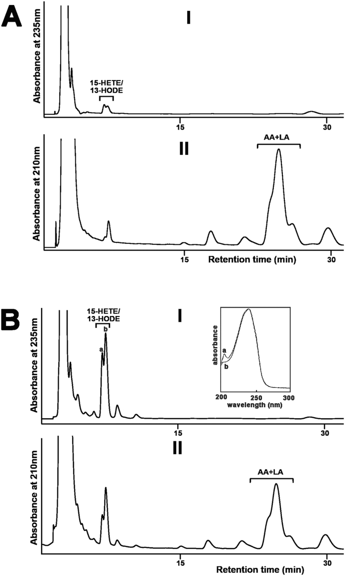
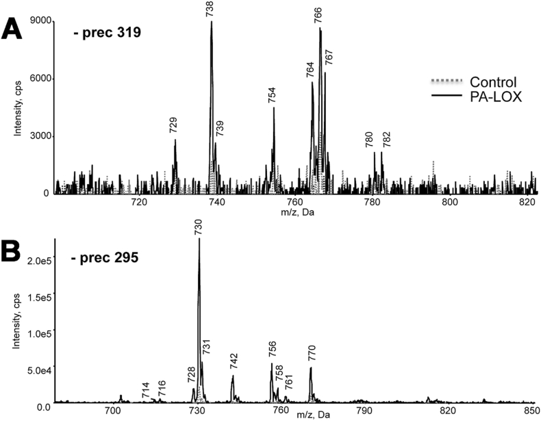
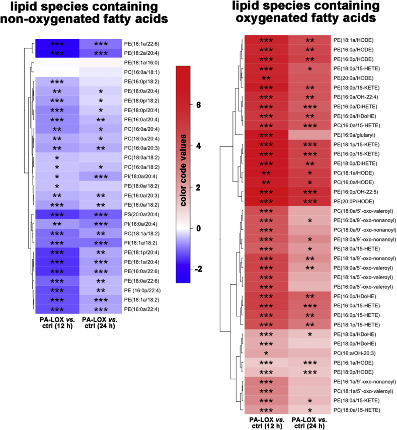
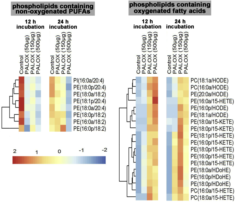
Pseudomonas aeruginosa is a gram-negative pathogen, which causes life-threatening infections in immunocompromized patients. These bacteria express a secreted lipoxygenase (PA-LOX), which oxygenates free arachidonic acid to 15S-hydro(pero)xyeicosatetraenoic acid. It binds phospholipids at its active site and physically interacts with lipid vesicles. When incubated with red blood cells membrane lipids are oxidized and hemolysis is induced but the structures of the oxygenated membrane lipids have not been determined. Using a lipidomic approach, we analyzed the formation of oxidized phospholipids generated during the in vitro incubation of recombinant PA-LOX with human erythrocytes and cultured human lung epithelial cells. Precursor scanning of lipid extracts prepared from these cells followed by multiple reaction monitoring and MS/MS analysis revealed a complex mixture of oxidation products. For human red blood cells this mixture comprised forty different phosphatidylethanolamine and phosphatidylcholine species carrying oxidized fatty acid residues, such as hydroxy-octadecadienoic acids, hydroxy- and keto-eicosatetraenoic acid, hydroxy-docosahexaenoic acid as well as oxygenated derivatives of less frequently occurring polyenoic fatty acids. Similar oxygenation products were also detected when cultured lung epithelial cells were employed but here the amounts of oxygenated lipids were smaller and under identical experimental conditions we did not detect major signs of cell lysis. However, live imaging indicated an impaired capacity for trypan blue exclusion and an augmented mitosis rate. Taken together these data indicate that PA-LOX can oxidize the membrane lipids of eukaryotic cells and that the functional consequences of this reaction strongly depend on the cell type.

Keywords: Biomembranes; Eicosanoids; Fatty acids; Infectious diseases; Lipidomics; Oxidative stress; Phospholipids.


Figures

Fig. 1:
Fig. 1
Oxygenation of linoleic acid and arachidonic acid esterified in the membrane phospholipids of intact erythrocytes to 13-HODE and 15-HETE by purified PA-LOX. Purified PA-LOX (2 μg, 7 μg, 60 μg, 170 μg and 500 μg) was incubated with 100 μl of packed erythrocytes in 1 ml of PBS (pH 7.4) at 25 °C for 24 h under continuous agitation. Similar incubations of the erythrocytes were carried out in the absence of PA-LOX as non-enzyme controls. After 24 h, the reaction mixtures were subjected to lipid extraction. The bottom chloroform phase was recovered, the solvent was evaporated and the remaining lipids were reconstituted in methanol. The extracted lipids were then hydrolyzed under alkaline conditions and analyzed by RP-HPLC to determine the major oxygenation products formed in the A) non-enzymatic control and B) PA-LOX containing sample. Similar chromatograms were obtained for all PA-LOX concentrations (five samples) but only the chromatogram for the 500 μg incubation is shown. Inset: UV-spectra of the conjugated dienes taken at the chromatographic time points indicated by a and b.
Fig. 2:
Fig. 2
Precursor scanning of RBC lipid extracts identified numerous phospholipid species that are decreased following PA-LOX treatment. Human erythrocytes were isolated from healthy volunteers and 100 μl of packed erythrocytes were incubated in 1 ml PBS in the presence/absence of 385 μg/ml of PA-LOX for 12 h at 25 °C, followed by lipid extraction. LC/MS/MS showing precursor scans of control (dashed line) or PA-LOX treated (solid line) red blood cell lipid extracts. Spectra were acquired scanning Q1 from 650 to 950 amu, following direct infusion. Panel A. Identification of ions that lose 196 amu in negative mode, consistent with PE. Panel B. Identification of ions that lose 241 amu in negative mode, consistent with PI. Panel C. Identification of ions that lose 184 amu in positive mode, consistent with PC. Panel D. Identification of ions that lose 87 amu in negative mode, consistent with PS. Four different PA-LOX incubations and two independent no-enzyme controls were carried out. A representative precursor scan is shown.
Fig. 3:
Fig. 3
Precursor ion scanning of RBC lipid extracts identified several phospholipids containing linoleic acid and arachidonic acid that are decreased following PA-LOX treatment. Human erythrocytes were isolated from healthy volunteers and 100 μl of packed erythrocytes were incubated in 1 ml PBS in the presence/absence of 385 μg/ml of PA-LOX for 12 h at 25 °C, followed by lipid extraction. LC/MS/MS showing precursor scans of control (dashed line) or PA-LOX treated (solid line) RBC lipid extracts. Spectra were acquired scanning Q1 from 650 to 950 amu, following direct infusion. Panel A. Precursor scan of m/z 303.2 shows lipids containing arachidonic acid. Panel B. Precursor scan of m/z 279.2 shows lipids containing linoleic acid. The incubations were carried out in duplicate and each lipid extract analyzed twice. As indicated in the legend to Fig. 2 four different PA-LOX incubations and two independent no-enzyme controls were carried out. A representative precursor scan is shown.
Fig. 4:
Fig. 4
Precursor scanning of RBC lipid extracts revealed generation of HETE-PE/PC and HODE-PE/PC following PA-LOX treatment. Human erythrocytes were isolated from healthy volunteers and incubated in the presence/absence of 385 μg of PA-LOX for 12 h at 25 °C, followed by lipid extraction. LC/MS/MS showing precursor scans of control (dashed line) or PA-LOX treated (solid line) RBC lipid extracts. Spectra were acquired, in negative mode, scanning Q1 from 650 to 950 amu, following direct infusion. Panel A. Identification of ions that generate m/z 319.2 daughter ions. Panel B. Identification of ions that generate m/z 295.1 daughter ions. As indicated in the legend to Fig. 2 four different PA-LOX incubations and two independent no-enzyme controls were carried out. A representative precursor scan is shown.
Fig. 5:
Fig. 5
Effect of purified PA-LOX on phospholipid composition of human erythrocytes, following 12 and 24-h in vitro incubation. Heatmaps were generated using ratio analyte to internal standard data using the pheatmap package in R. Levels of treatment response are represented by a color gradient ranging from blue (decrease in response) to white (no change) to red (increase in response). Lipids are color-coded by group and clustered by similarity in overall response to PA-LOX. Lipids were normalized to basal levels, at 12 h. As indicated in the legend to Fig. 2 four different PA-LOX incubations and two independent no-enzyme controls were carried out.
Fig. 6
Fig. 6
Alterations in the concentrations of different lipid species during PA-LOX treatment of red blood cell membranes. The color code semi-quantitatively mirrors the alterations in lipid concentrations. Tukey's Honestly Significant Differences post-hoc test was used to compare two groups after one-way analysis of variance. Blue color represents a decrease in the concentration of a particular lipid species and red color indicates an increase. The significances of the differences (*p < 0.05, **p < 0.005, ***p < 0.0005; t-test) are marked. As indicated in the legend to Fig. 2 four different PA-LOX incubations and two independent no-enzyme controls were carried out.
Fig. 7:
Fig. 7
Analysis of selected phospholipid species formed when A549 cells were treated with purified PA-LOX. A549 cells were incubated in the presence/absence of varying amounts of PA-LOX (50 μg, 150 μg, 500 μg/ml) for 12 h and 24 h. Lipid extracts were analyzed by reverse-phase LC/MS/MS, in negative mode, using Luna column on 6500 Q Trap. Single incubations were carried out for each PA-LOX concentration. Each sample was analyzed once by RP-HPLC and in triplicate LC-MS/MS.
Fig. 8:
Fig. 8
Confocal light micrographs of cultured A549 cells. Pre-confluenet A549 cells were incubated in the absence (panel A) and presence (panel B) of 250 μg/ml pure recombinant PA-LOX for 16 h. Four different no-enzyme controls wells and four PA-LOX incubation wells were set up. Representative images are shown and mitotic cells are labeled by the asterix.
Fig. 9:
Fig. 9
Correlation analysis of different PA-LOX substrate lipids present in erythrocyte membranes. A) Correlations among fold changes of different PA-LOX substrate lipids after treatment of human erythrocytes for 12 h in the presence and absence of PA-LOX. B) Correlations among fold changes of different PA-LOX substrate lipids after 12 h and 24 h treatment of human erythrocytes in the absence of PA-LOX. Red color indicates strong positive correlation; the brighter the red color, the stronger positive correlations. Blue color indicates strong negative correlation; the brighter the blue color, the stronger negative correlations. The area of each circle symbolizes the absolute value of its corresponding correlation. As indicated in the legend to Fig. 2 four different PA-LOX incubations and two independent no-enzyme controls were carried out. A representative precursor scan is shown.

Similar articles

Cited by

References

    1. Andreou A., Brodhun F., Feussner I. Biosynthesis of oxylipins in non-mammals. Prog. Lipid Res. 2009;48:148–170. - PubMed
    1. Kuhn H., Banthiya S., van Leyen K. Mammalian lipoxygenases and their biological relevance. Biochim. Biophys. Acta. 2015;1851:308–330. - PMC - PubMed
    1. Haeggstrom J.Z., Funk C.D. Lipoxygenase and leukotriene pathways: biochemistry, biology, and roles in disease. Chem. Rev. 2011;111:5866–5898. - PubMed
    1. de Bentzmann S., Plesiat P. The Pseudomonas aeruginosa opportunistic pathogen and human infections. Environ. Microbiol. 2011;13:1655–1665. - PubMed
    1. Vance R.E., Hong S., Gronert K., Serhan C.N., Mekalanos J.J. The opportunistic pathogen Pseudomonas aeruginosa carries a secretable arachidonate 15-lipoxygenase. Proc. Natl. Acad. Sci. U. S. A. 2004;101:2135–2139. - PMC - PubMed

Publication types

LinkOut - more resources