Role of MR Neurography for the Diagnosis of Peripheral Trigeminal Nerve Injuries in Patients with Prior Molar Tooth Extraction
- PMID: 29146720
- PMCID: PMC7410708
- DOI: 10.3174/ajnr.A5438
Role of MR Neurography for the Diagnosis of Peripheral Trigeminal Nerve Injuries in Patients with Prior Molar Tooth Extraction
Abstract
Background and purpose: Clinical neurosensory testing is an imperfect reference standard to evaluate molar tooth extraction related peripheral trigeminal neuropathy. The purpose was to evaluate the diagnostic accuracy of MR neurography in this domain and correlation with neurosensory testing and surgery.
Materials and methods: In this retrospective study, nerve caliber, T2 signal intensity ratio, and contrast-to-noise ratios were recorded by 2 observers using MR neurography for bilateral branches of the peripheral trigeminal nerve, the inferior alveolar and lingual nerves. Patient demographics and correlation of the MR neurography findings with the Sunderland classification of nerve injury and intraoperative findings of surgical patients were obtained.
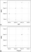
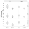
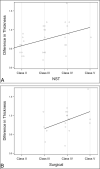
Results: Among 42 patients, the mean ± SD age for case and control patients were 35.8 ± 10.2 years and 43.2 ± 11.5 years, respectively, with male-to-female ratios of 1:1.4 and 1:5, respectively. Case subjects (peripheral trigeminal neuropathy or injury) had significantly larger differences in nerve thickness, T2 signal intensity ratio, and contrast-to-noise ratios than control patients for the inferior alveolar nerve and lingual nerve (P = .01 and .0001, .012 and .005, and .01 and .01, respectively). Receiver operating characteristic analysis showed a significant association among differences in nerve thickness, T2 signal intensity ratio, and contrast-to-noise ratios and nerve injury (area under the curve, 0.83-0.84 for the inferior alveolar nerve and 0.77-0.78 for the lingual nerve). Interobserver agreement was good for the inferior alveolar nerve (intraclass correlation coefficient, 0.70-0.79) and good to excellent for the lingual nerve (intraclass correlation coefficient, 0.75-0.85). MR neurography correlations with respect to clinical neurosensory testing and surgical classifications were moderate to good. Pearson correlation coefficients of 0.68 and 0.81 and κ of 0.60 and 0.77 were observed for differences in nerve thickness.
Conclusions: MR neurography can be reliably used for the diagnosis of injuries to the peripheral trigeminal nerve related to molar tooth extractions, with good to excellent correlation of imaging with clinical findings and surgical results.
© 2018 by American Journal of Neuroradiology.
Figures


References
-
- Pogrel MA, Kaban LB. Injuries to the inferior alveolar and lingual nerves. J Calif Dent Assoc 1993;21:50–54 - PubMed
-
- American Dental Association. 1999 survey of dental services rendered. ADA Catalog No SDSR-1999
Publication types
MeSH terms
LinkOut - more resources
Full Text Sources
Other Literature Sources
Medical
