. 2018 Mar;32(3):1537-1549.
doi: 10.1096/fj.201700740R. Epub 2018 Jan 3.

Humanized mice in studying efficacy and mechanisms of PD-1-targeted cancer immunotherapy

Affiliations

Humanized mice in studying efficacy and mechanisms of PD-1-targeted cancer immunotherapy

Minan Wang et al. FASEB J. 2018 Mar.

Abstract

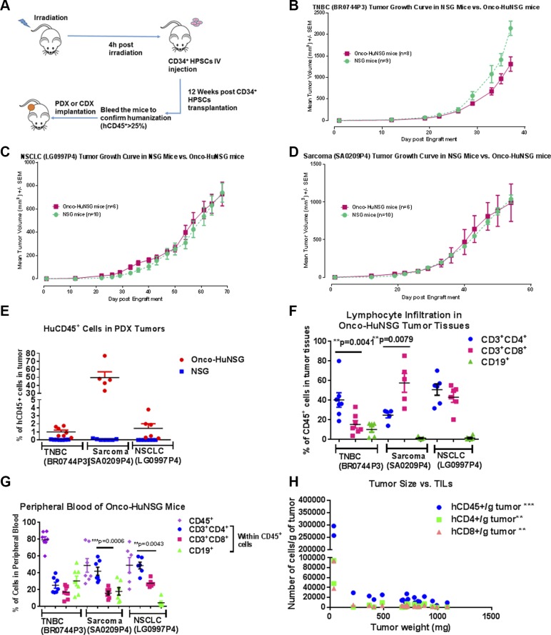
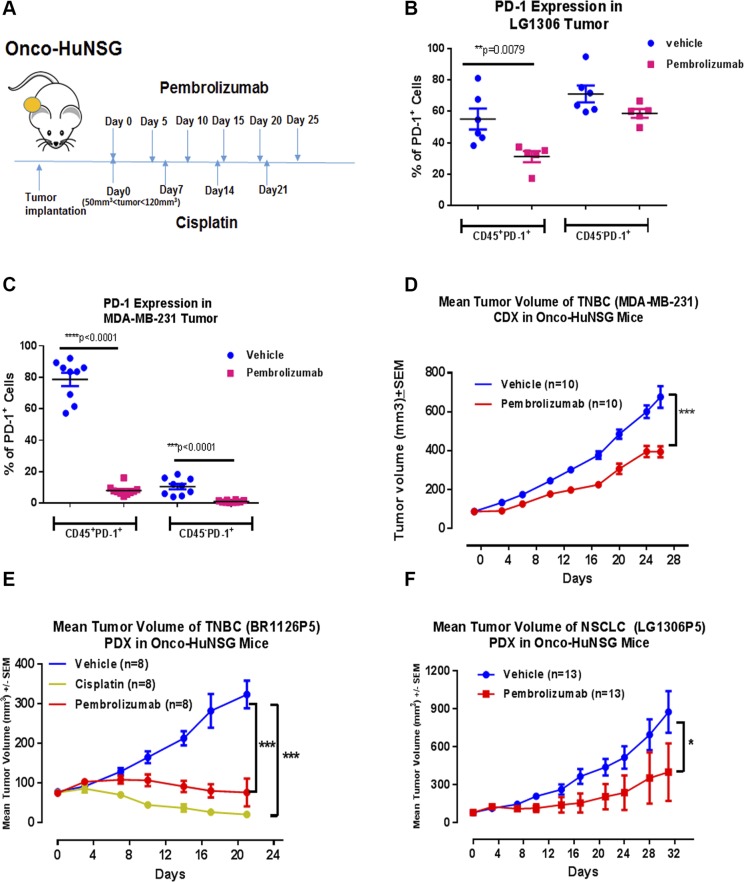
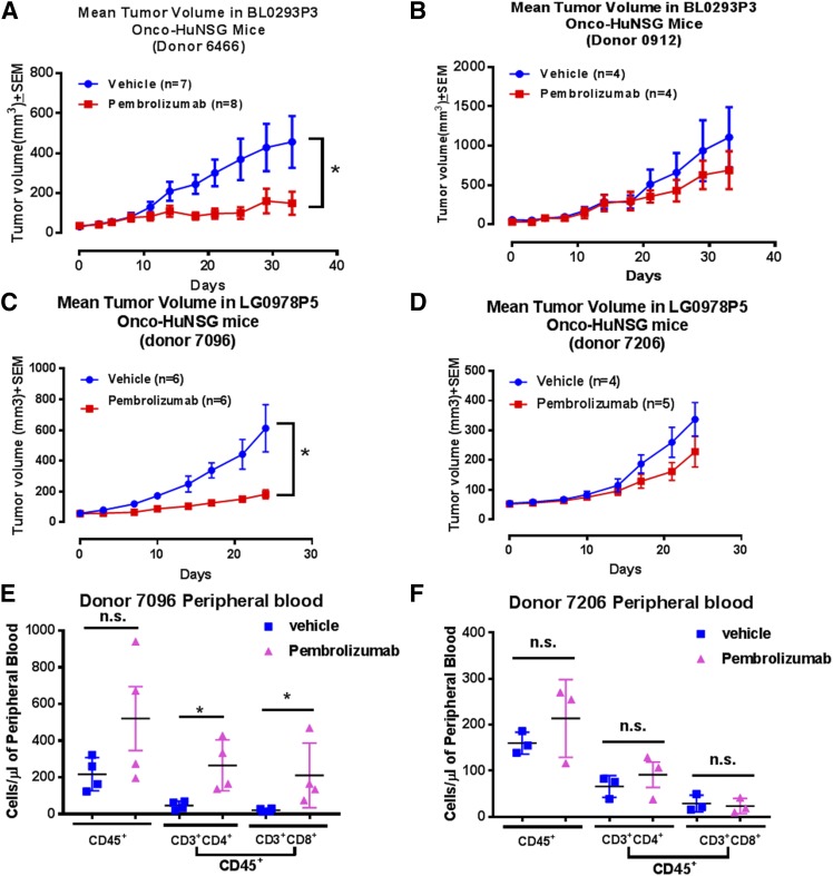
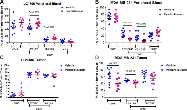
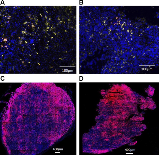
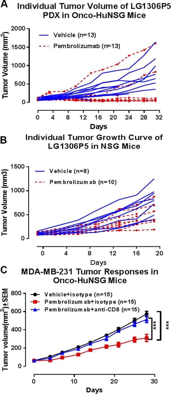
Establishment of an in vivo small animal model of human tumor and human immune system interaction would enable preclinical investigations into the mechanisms underlying cancer immunotherapy. To this end, nonobese diabetic (NOD).Cg- PrkdcscidIL2rgtm1Wjl/Sz (null; NSG) mice were transplanted with human (h)CD34+ hematopoietic progenitor and stem cells, which leads to the development of human hematopoietic and immune systems [humanized NSG (HuNSG)]. HuNSG mice received human leukocyte antigen partially matched tumor implants from patient-derived xenografts [PDX; non-small cell lung cancer (NSCLC), sarcoma, bladder cancer, and triple-negative breast cancer (TNBC)] or from a TNBC cell line-derived xenograft (CDX). Tumor growth curves were similar in HuNSG compared with nonhuman immune-engrafted NSG mice. Treatment with pembrolizumab, which targets programmed cell death protein 1, produced significant growth inhibition in both CDX and PDX tumors in HuNSG but not in NSG mice. Finally, inhibition of tumor growth was dependent on hCD8+ T cells, as demonstrated by antibody-mediated depletion. Thus, tumor-bearing HuNSG mice may represent an important, new model for preclinical immunotherapy research.-Wang, M., Yao, L.-C., Cheng, M., Cai, D., Martinek, J., Pan, C.-X., Shi, W., Ma, A.-H., De Vere White, R. W., Airhart, S., Liu, E. T., Banchereau, J., Brehm, M. A., Greiner, D. L., Shultz, L. D., Palucka, K., Keck, J. G. Humanized mice in studying efficacy and mechanisms of PD-1-targeted cancer immunotherapy.

Keywords: checkpoint inhibitor; mouse model; patient-derived xenograft; pembrolizumab.


Conflict of interest statement

The authors thank the Flow Contract Site Laboratory (Bothell, WA, USA) for processing and staining samples for flow cytometry, and Pure Transplant Solutions (Oklahoma City, OK, USA) for HLA typing. This study was funded by The Jackson Laboratory. The authors declare no conflicts of interest.

Figures

Figure 1.
Figure 1.
HuNSG mice support growth of partially HLA-matched human CDX and PDX tumors and immune cell populations in Onco-HuNSG mice. A) Experimental design for generating HuNSG mice bearing CDX or PDX tumors. B–D) Implantation of HLA partially matched PDX tumors BR0744P3 (B), LG0997P4 (C), and SA0209P4 (D) into NSG or HuNSG mice. PDX tumors were excised and implanted subcutaneously into NSG or HuNSG mice. Tumor measurements were calculated as (L × W × W)/2, where L is the length, and W is the width of the tumor. No statistical differences were observed in any tumor model between NSG and HuNSG mice. E) Flow cytometric analysis of hCD45+ cells in tumors from NSG or HuNSG mice. No hCD45+ cells were detected in tumors growing in NSG mice. F, G) At the study end point, hCD3, hCD4, hCD8, and hCD19 were stained for flow cytometric analysis to determine the populations of lymphocytes in tumor tissues (F) and peripheral blood (G) of 3 different PDX models. CD3+, CD4+, CD19+ cells are presented as percentage of cells within CD45+ cells. H) hCD45, hCD3, hCD4, and hCD8 were stained for flow cytometric analysis to determine numbers of TILs in MDA-MB-231 tumor-bearing HuNSG mice. Correlation was calculated by GraphPad Prism 5. **P < 0.01, ***P < 0.001.
Figure 2.
Figure 2.
Pembrolizumab inhibited tumor growth in Onco-HuNSG mice. A) Experimental design and treatment schedule for anti-PD-1 and chemotherapy against CDX and PDX tumors. Treatments started when tumors reached 50–120 mm3 in volume. B, C) Animals were randomized into different experimental groups. Tumors from LG1306P5 (B) and MDA-MB-231 (C) Onco-HuNSG mice were processed into single-cell suspensions and stained, as described in Materials and Methods, for flow cytometric analysis. PD-1 was stained on both CD45 and CD45+ populations. D) MDA-MB-231 human breast cancer cells (5 × 10⁶) in matrigel were injected into mammary fat pads of HuNSG mice. Vehicle control saline was injected intraperitoneally every 5 d for 25 d. Pembrolizumab was injected at 10 mg/kg on d 0, followed by 5 mg/kg on d 5, 10, 15, 20, and 25. E) TNBC PDX tumor BR1126P5 was trocared subcutaneously into HuNSG mice. Vehicle control saline was injected intraperitoneally every 5 d for 25 d. Cisplatin was injected at 2 mg/kg, i.v. on d 0, 7, and 14. Pembrolizumab was injected at 10 mg/kg, i.p. on d 0, followed injection at 5 mg/kg, i.p. on d 5, 10, 15, and 20. On d 21, both the cisplatin and pembrolizumab treatment groups have significantly smaller tumor size compared with the vehicle control group. F) NSCLC PDX LG1306P5 was trocared subcutaneously into HuNSG mice. Vehicle control saline and pembrolizumab (5 mg/kg) were injected intraperitoneally every 5 d for 25 d. For experiments with 2 groups, tumor growth response curves were analyzed by the Multivariate ANOVA test, followed by univariate test with JMP 11 software (D, F). For the experiment with 3 groups, tumor growth curves were analyzed by 2-way ANOVA, followed by Dunnett’s posttests using GraphPad Prism (E). *P < 0.05, **P < 0.01, ***P < 0.001.
Figure 3.
Figure 3.
Donor variations in response to pembrolizumab treatment in bladder (BL0293P3) and NSCLC (LG0978P5) Onco-HuNSG mice. A, B) Tumor growth curves of PDX bladder tumor BL0293P3 Onco-HuNSG in responder and nonresponder donors. The BL0293P3 tumor was implanted subcutaneously into HuNSG mice, established from 2 donor CD34+ HPSC sources: donor 6466 (A) and donor 0912 (B). Vehicle control saline was injected intraperitoneally twice a week until the end of the study. Pembrolizumab was injected 10 mg/kg, i.p. on d 0 followed by 5 mg/kg twice a week until the end of the study. C, D) Tumor growth curves of PDX NSCLC (LG0978P5) Onco-HuNSG in responder and nonresponder donors. The LG0978P5 tumor was trocared subcutaneously into HuNSG mice from 2 donor CD34+ HPSC sources: donor 7096 (C) and donor 7206 (D). Vehicle control saline was injected intraperitoneally every 5 d for 20 d on d 0, 5, 10, 15, and 20. Pembrolizumab was injected at 10 mg/kg, i.p. on d 0, followed by 5 mg/kg, i.p. injection on d 5, 10, 15, and 20. E, F) Whole blood from NSCLC (LG0978P5) Onco-HuNSG donor 7096 (E) and donor 7206 (F) mice was processed into single-cell suspension and stained for hCD45, hCD3, hCD4, and hCD8, as described in Materials and Methods, for flow cytometric analysis. Nonparametric Mann-Whitney U test was performed to compare the cell numbers in different treatment groups. *P < 0.05.
Figure 4.
Figure 4.
Human immune cell populations in peripheral blood and tumors of the NSCLC (LG1306) and TNBC (MDA-MB-231) Onco-HuNSG model. Whole blood from LG1306 (A) or MDA-MB-231 (B) Onco-HuNSG mice was processed into single-cell suspensions and stained, as described in Materials and Methods, for flow cytometric analysis. Tumor tissues from LG1306 (C) or MDA-MB-231 (D) Onco-HuNSG mice were processed into single-cell suspensions and stained, as described in Materials and Methods, for flow cytometric analysis. The percentage of hCD3+CD4+ T cells, hCD3+CD8+ T cells, and hCD19+ B cells is shown within CD45+ cells. *P < 0.05; **P < 0.01.
Figure 5.
Figure 5.
In situ characterization of CD8+ T-cell infiltration into the tumor. A, B) Tumor tissues from LG1306 were processed for histologic analysis and triple stained with DAPI (blue), hCD45 (yellow), and hCD8 (red). Representative tumor samples are shown from LG1306 treated with vehicle control (A) or pembrolizumab (B). CD8+ T cells were observed in both the tumor margin and center. C, D) Tumor tissues from MDA-MB-231 were processed for histologic analysis and stained with DAPI (blue) and hCD8 (red). Representative tumor samples are shown from MDA-MB-231 treated with vehicle control (C) or pembrolizumab (D). More CD8+ T cells were observed in the tumor center than the tumor margin in the pembrolizumab-treated tumor.
Figure 6.
Figure 6.
Efficacy of pembrolizumab is mediated by hCD8+ T cells in Onco-HuNSG mice. NSCLC PDX LG1306P5 was implanted in HuNSG mice (A) or NSG mice (B). A) Vehicle control saline or pembrolizumab (5 mg/kg) was injected intraperitoneally every 5 d for 30 d on d 0, 5, 10, 15, 20, and 25 in HuNSG mice. B) Vehicle control saline and pembrolizumab (5 mg/kg) was injected intraperitoneally every 5 d for 20 d on d 0, 5, 10, and 15 in NSG mice. C) MDA-MB-231 cells (5 × 10⁶) were injected into the mammary fat pads in HuNSG mice. When the tumor reached 50–120 mm3, the mice were treated with pembrolizumab or vehicle on d 1, 6, 11, 16, 21, and 26. Isotype control antibody or anti-hCD8 antibody was injected intraperitoneally on d 0, 7, 14, 21, and 28. Tumor growth response curves were analyzed by 2-way ANOVA, followed by Dunnett’s posttests. ***P < 0.001.

References

    1. Hackam D. G., Redelmeier D. A. (2006) Translation of research evidence from animals to humans. JAMA 296, 1731–1732 https://doi.org/10.1001/jama.296.14.1731 - DOI - PubMed
    1. Mestas J., Hughes C. C. (2004) Of mice and not men: differences between mouse and human immunology. J. Immunol. 172, 2731–2738 https://doi.org/10.4049/jimmunol.172.5.2731 - DOI - PubMed
    1. Theocharides A. P., Rongvaux A., Fritsch K., Flavell R. A., Manz M. G. (2016) Humanized hemato-lymphoid system mice. Haematologica 101, 5–19 https://doi.org/10.3324/haematol.2014.115212 - DOI - PMC - PubMed
    1. Rongvaux A., Willinger T., Martinek J., Strowig T., Gearty S. V., Teichmann L. L., Saito Y., Marches F., Halene S., Palucka A. K., Manz M. G., Flavell R. A. (2014) Development and function of human innate immune cells in a humanized mouse model. Nat. Biotechnol. 32, 364–372 https://doi.org/10.1038/nbt.2858 - DOI - PMC - PubMed
    1. Ishikawa F., Yasukawa M., Lyons B., Yoshida S., Miyamoto T., Yoshimoto G., Watanabe T., Akashi K., Shultz L. D., Harada M. (2005) Development of functional human blood and immune systems in NOD/SCID/IL2 receptor gamma chain(null) mice. Blood 106, 1565–1573 https://doi.org/10.1182/blood-2005-02-0516 - DOI - PMC - PubMed

Publication types

Substances