. 2018 Jan;176(1):418-431.
doi: 10.1104/pp.17.01075. Epub 2017 Nov 16.

Division Plane Orientation Defects Revealed by a Synthetic Double Mutant Phenotype

Affiliations

Division Plane Orientation Defects Revealed by a Synthetic Double Mutant Phenotype

Ricardo Mir et al. Plant Physiol. 2018 Jan.

Abstract

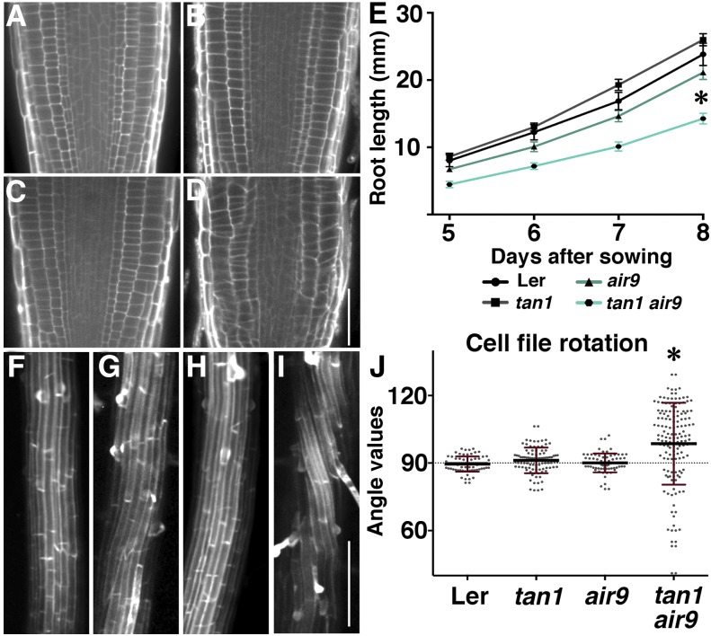
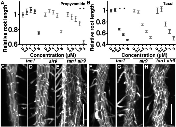
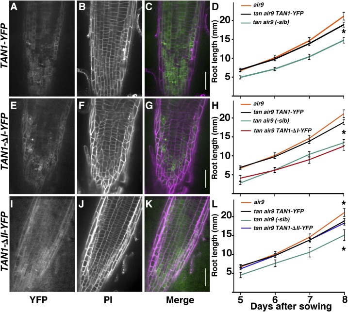
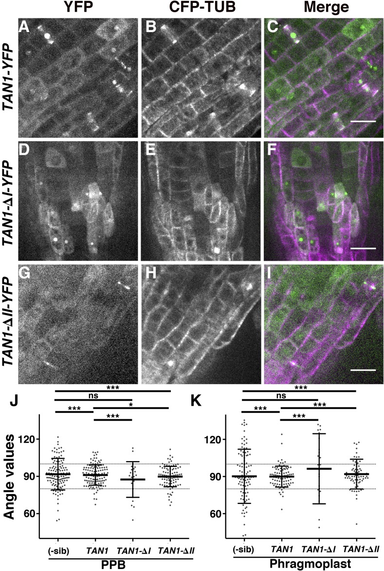
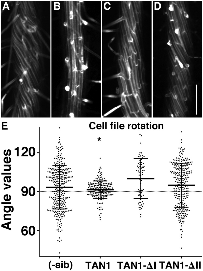
TANGLED1 (TAN1) and AUXIN-INDUCED-IN-ROOTS9 (AIR9) are microtubule-binding proteins that localize to the division site in plants. Their function in Arabidopsis (Arabidopsis thaliana) remained unclear because neither tan1 nor air9 single mutants have a strong phenotype. We show that tan1 air9 double mutants have a synthetic phenotype consisting of short, twisted roots with disordered cortical microtubule arrays that are hypersensitive to a microtubule-depolymerizing drug. The tan1 air9 double mutants have significant defects in division plane orientation due to failures in placing the new cell wall at the correct division site. Full-length TAN1 fused to yellow fluorescent protein, TAN1-YFP, and several deletion constructs were transformed into the double mutant to assess which regions of TAN1 are required for its function in root growth, root twisting, and division plane orientation. TAN1-YFP expressed in tan1 air9 significantly rescued the double mutant phenotype in all three respects. Interestingly, TAN1 missing the first 126 amino acids, TAN1-ΔI-YFP, failed to rescue the double mutant phenotype, while TAN1 missing a conserved middle region, TAN1-ΔII-YFP, significantly rescued the mutant phenotype in terms of root growth and division plane orientation but not root twisting. We use the tan1 air9 double mutant to discover new functions for TAN1 and AIR9 during phragmoplast guidance and root morphogenesis.


Figures

Figure 1.
Figure 1.
Root phenotypes of Ler, tan1, air9, and tan1 air9 double mutants. A to D, Root cell walls stained with propidium iodide of Ler (A), tan1 (B), air9 (C), and tan1 air9 (D) plants. E, Root length measurements from days 5 to 8 after sowing of Ler and mutant plants onto vertically oriented plates. The asterisk indicates that tan1 air9 roots were significantly shorter than others (P < 0.01). F to I, Maximum projections of 10 0.2-µm Z-stacks showing expansion to the differentiation zone of propidium iodide-stained roots of Ler (F), tan1 (G), air9 (H), and tan1 air9 (I). J, Root cell file rotation (twisting) measurements. Each dot represents an angle measured from the transverse wall to the long axis of the root. Angles greater than 90° are left twisting and angles less than 90° are right twisting. The asterisk indicates a significant difference in the distribution of tan1 air9 cell file rotation compared with others (P < 0.01). Bars = 50 µm (A–D) and 200 µm (F–I).
Figure 2.
Figure 2.
Effects of propyzamide and taxol on tan1, air9, and tan1 air9 double mutant plants. A and B, Relative root length normalized by lengths of the 0 µm-treated roots of 8-d-old tan1, air9, and tan1 air9 plants grown on different concentrations of propyzamide (A) and taxol (B). Asterisks indicate significantly different responses to drug treatments (Kolmogorov–Smirnov test, P < 0.01; n > 24 plants per condition each day). C to H, Maximum projections of 10 (C, D, and F–H) and 30 (E) 1-µm Z-stacks of 8-d-old propidium iodide-stained differentiation zone roots of tan1 (C and F), air9 (D and G), and tan1 air9 (E and H) plants treated with 3 µm propyzamide (C–E) and 3 µm taxol (F–H). Bars = 200 µm.
Figure 3.
Figure 3.
PPB and phragmoplast angle measurements, micrographs, and time-lapse imaging of dividing cells expressing CFP-TUBULIN. A, PPB and phragmoplast angle orientation in dividing root cells. The angle was measured between the long axis of the cell wall and the orientation of the PPB or phragmoplast. The 80° and 100° angles are indicated by dotted lines. A line heterozygous for air9 and tan1 in the Ler background was used for Ler. Asterisks indicate statistically significant differences in distributions (F test, P < 0.0001). B and C, Merged confocal images showing CFP-TUBULIN (green) and propidium iodide (magenta) of tan1 air9 double mutant root cells. Images show PPBs (B) and phragmoplasts (C) with angles outside the 80° to 100° range. D to K, Time-lapse images of tan1 (D), air9 (F), oriented tan1 air9 (H), and misoriented tan1 air9 (J) division, showing the different phases of mitosis. Merged images of completed division starting with a PPB (green) and ending with a new cell wall (magenta) are shown for tan1 (E), air9 (G), properly oriented tan1 air9 division (I), and misoriented cell wall in a tan1 air9 division (K). Minutes and seconds are given in white at the bottom left sides of the time-lapse images. Bars = 5 µm.
Figure 4.
Figure 4.
Root micrographs and root length measurements of tan1 air9 plants expressing full-length TAN1-YFP, TAN1-∆I-YFP, and TAN1-∆II-YFP. A to C, E to G, and I to K, Confocal images of root cell walls stained with propidium iodide (PI) of tan1 air9 plants expressing TAN1-YFP (A–C), TAN1-∆I-YFP (E–G), and TAN1-∆II-YFP (I–K) with YFP signal (A, E, and I), cell walls stained with PI (B, F, and J), and merged channels (C, G, and K). YFP signal is green and PI signal is magenta. Bars = 50 µm. D, H, and L, Root length measurements from day 5 to 8 of tan1 air9 plants expressing full-length TAN1-YFP (D), TAN1-∆I-YFP (H), and TAN1-∆II-YFP (L). Single mutant air9 and nontransformed sibling plants were used as controls. Asterisks indicate significant differences between root lengths by Student’s t test (P < 0.01).
Figure 5.
Figure 5.
PPB and phragmoplast orientation of tan1 air9 double mutants expressing full-length TAN1-YFP, TAN1-∆I-YFP, and TAN1-∆II-YFP. A to I, Confocal images of propidium iodide-stained roots of tan1 air9 plants with CFP-TUBULIN and TAN1-YFP (A–C), TAN1-∆I-YFP (D–F), and TAN1-∆II-YFP (G–I). Shown are YFP signal (A, D, and G), CFP-TUBULIN signal (B, E, and H), and merged images (C, F, and I). Bars = 20 µm. J and K, Orientation compared with the lateral cell walls of PPBs (J) and phragmoplasts (K) of tan1 air9 plants coexpressing CFP-TUBULIN and YFP-tagged versions of full-length TAN1, TAN1-∆I, and TAN1-∆II, as indicated. YFP-negative sibling plants (-sib) were used to measure the orientation of PPBs and phragmoplasts as a control. Each dot represents a measured angle. The F test was used to compare distributions. The single asterisk indicates P = 0.02, triple asterisks indicate P < 0.002, and ns indicates no significant difference detected.
Figure 6.
Figure 6.
Differentiation zones of tan1 air9 roots expressing full-length TAN1-YFP, TAN1-∆I-YFP, and TAN1-∆II-YFP. A to D, Maximum projections of 10 Z-stacks of 0.2 µm of propidium iodide-stained roots of tan1 air9 plants (A) and tan1 air9 plants expressing TAN1-YFP (B), TAN1-∆I-YFP (C), and TAN1-∆II-YFP (D). Bar = 200 µm. E, Cell file rotation angles of double mutant tan1 air9 plants expressing TAN1-YFP, TAN1-∆I-YFP, or TAN1-∆II-YFP and the corresponding nontransformed sibling plants (-sib). Angles greater than 90° are left twisting and angles less than 90° are right twisting. The asterisk indicates a significantly smaller variance (P < 0.001 by Levene’s test; n = 330).

Similar articles

Cited by

References

    1. Abrash EB, Bergmann DC (2009) Asymmetric cell divisions: a view from plant development. Dev Cell 16: 783–796 - PubMed
    1. Ambrose JC, Cyr R (2008) Mitotic spindle organization by the preprophase band. Mol Plant 1: 950–960 - PubMed
    1. Azimzadeh J, Nacry P, Christodoulidou A, Drevensek S, Camilleri C, Amiour N, Parcy F, Pastuglia M, Bouchez D (2008) Arabidopsis TONNEAU1 proteins are essential for preprophase band formation and interact with centrin. Plant Cell 20: 2146–2159 - PMC - PubMed
    1. Baskin TI. (2001) On the alignment of cellulose microfibrils by cortical microtubules: a review and a model. Protoplasma 215: 150–171 - PubMed
    1. Besson S, Dumais J (2014) Stochasticity in the symmetric division of plant cells: when the exceptions are the rule. Front Plant Sci 5: 538. - PMC - PubMed

Publication types

MeSH terms

LinkOut - more resources