. 2017 Nov 17;358(6365):888-893.
doi: 10.1126/science.aao1140.

The structural basis of flagellin detection by NAIP5: A strategy to limit pathogen immune evasion

Affiliations

The structural basis of flagellin detection by NAIP5: A strategy to limit pathogen immune evasion

Jeannette L Tenthorey et al. Science. .

Abstract

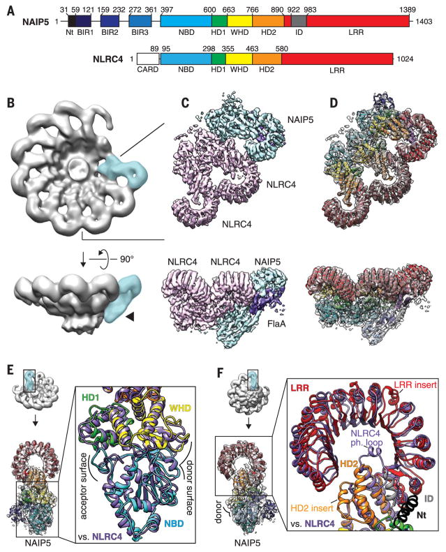
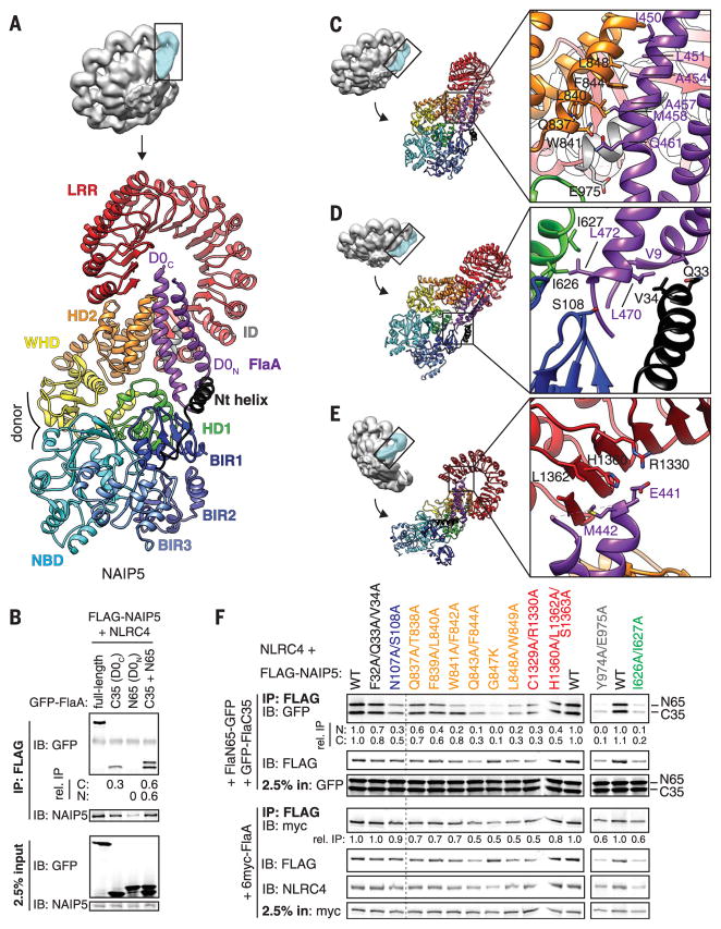
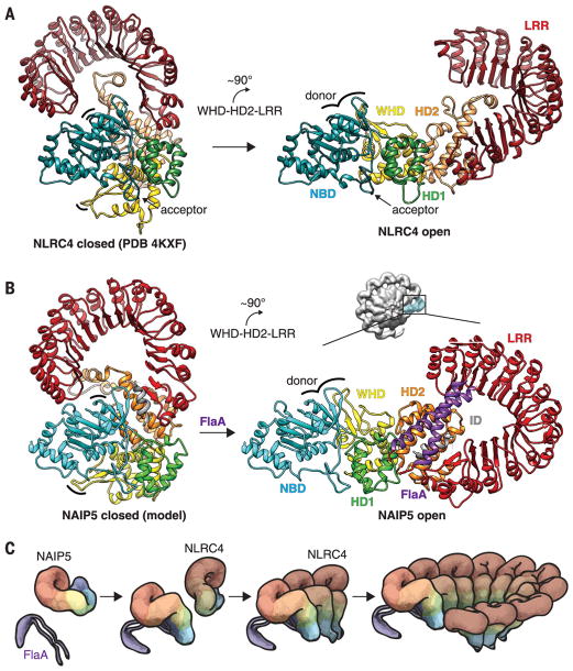
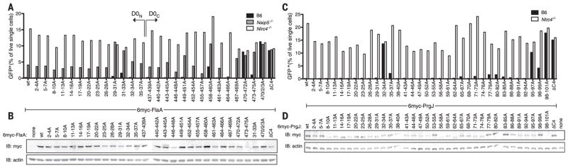
Robust innate immune detection of rapidly evolving pathogens is critical for host defense. Nucleotide-binding domain leucine-rich repeat (NLR) proteins function as cytosolic innate immune sensors in plants and animals. However, the structural basis for ligand-induced NLR activation has so far remained unknown. NAIP5 (NLR family, apoptosis inhibitory protein 5) binds the bacterial protein flagellin and assembles with NLRC4 to form a multiprotein complex called an inflammasome. Here we report the cryo-electron microscopy structure of the assembled ~1.4-megadalton flagellin-NAIP5-NLRC4 inflammasome, revealing how a ligand activates an NLR. Six distinct NAIP5 domains contact multiple conserved regions of flagellin, prying NAIP5 into an open and active conformation. We show that innate immune recognition of multiple ligand surfaces is a generalizable strategy that limits pathogen evolution and immune escape.


Figures

Fig. 1
Fig. 1. Structure of the NAIP5-NLRC4 inflammasome
(A) Schematic of domain architecture for NAIP5 and NLRC4. NAIP5 domains were defined in this work (methods); NLRC4 domains were previously defined (19). Residue numbers are shown. Nt, N terminus. (B) 3D reconstruction of inflammasomes containing a single NAIP5-FlaA (blue) and nine NLRC4 protomers (gray). The arrowhead highlights extra density that identified the NAIP5 protomer. (C) Refined 3D reconstruction for NAIP5 and the first two NLRC4 protomers at higher resolution (NAIP5 segmented in blue, the two NLRC4 protomers in pink, and FlaA in purple). (D) Modeled structures of NAIP5 and two NLRC4 protomers, all colored by domains as in (A), fitted within the EM map. In (B) to (D), two orthogonal views are shown. (E and F) The structure of NAIP5 [colored as in (A)] is aligned with that of an NLRC4 protomer (purple). (E) The NBD, HD1, and WHD oligomerization domains of NAIP5 and NLRC4 are highly similar. Oligomerization donor and acceptor surfaces [(9, 10) and fig. S5] are indicated. For clarity, the NAIP5 BIR domains were omitted from the inset view (right). (F) The HD2 and LRR of NAIP5 diverge from those of NLRC4. NAIP5-specific insertions, including an extra leucine-rich repeat (LRR insert; residues 1102 to 1138) and the modeled helix of the inserted domain (ID; gray), are indicated. The NLRC4 S533 phosphorylation loop is replaced by two alpha helices in NAIP5 (HD2 insert; residues 818 to 851).
Fig. 2
Fig. 2. Multiple NAIP5 domains contact extended surfaces on both helices of the flagellin D0 domain
(A) The D0 domain (purple) is locked into place by multiple NAIP5 domains. (B) Both D0 helices bind to NAIP5. (C to E) Detailed interactions between the flagellin D0 helices and the NAIP5 domains HD2 and ID (C); Nt, BIR1, and HD1 (D); and LRR (E). Side chains shown correspond to mutated residues in (F). (F) Mutagenesis confirms the importance of NAIP5 residues in binding D0N, D0C, or full-length FlaA. Point mutations in NAIP5 residues that contact D0C disrupt both D0N and D0C binding because association of D0N requires D0C binding [(B) and fig. S9A]. In (B) and (F), relative IP strength was quantified by densitometry [IP signal normalized to input and then GFP-FlaA (B) or WT NAIP5 (F)]. Results are representative of at least three independent experiments. IP, immunoprecipitation; IB, immunoblot; WT, wild type. Single-letter abbreviations for the amino acid residues are as follows: A, Ala; C, Cys; D, Asp; E, Glu; F, Phe; G, Gly; H, His; I, Ile; K, Lys; L, Leu; M, Met; N, Asn; P, Pro; Q, Gln; R, Arg; S, Ser; T, Thr; V, Val; W, Trp; and Y, Tyr.
Fig. 3
Fig. 3. Model of NAIP5-NLRC4 inflammasome assembly
(A) Structures of inactive (left) and active (right, determined in this work) NLRC4, showing a ~90° rigid-body rotation of the WHD-HD2-LRR module (9, 10) triggered by interaction with an activated NAIP5 or another already activated NLRC4. (B) To generate the inactive NAIP5 conformation [left; modeled based on NLRC4 (methods)], the HD2 insert was moved to avoid collision with the NBD. We propose that flagellin binding induces a ~90° rigid-body rotation of the WHD-HD2-LRR module, analogous to the rotation of NLRC4, which displaces the occluding LRR and HD2 from the NBD to complete and expose the donor oligomerization surface (indicated by curved lines in left and right panels) for interaction with NLRC4. (C) Proposed events of inflammasome assembly. The flagellin D0 domain (purple) binds to NAIP5 and unfurls the protein for subsequent NLRC4 recruitment and activation. Active NLRC4 recruits further NLRC4 protomers for self-propagating oligomerization and completion of a caspase-1 recruitment platform. Colors are as in Fig. 1A.
Fig. 4
Fig. 4. Multisurface ligand recognition is common to NAIPs
(A) Full-length 6myc-tagged L. pneumophila FlaA, or variants with the indicated residues mutated to alanine, were transduced into BMMs by using a retrovirus marked with IRES (internal ribosomal entry site)–GFP.Transduction efficiency was assessed by flow cytometry for GFP expression at day 4. Failure to transduce B6 BMMs, as compared with transduction of Nlrc4−/− BMMs, is indicative of NAIP activation (5). Naip5−/− BMM responses to FlaA are NAIP6-dependent (22). (B) Transduced Nlrc4−/− BMM lysates were probed for FlaA expression by anti-myc IB. (C) Full-length 6myc-tagged S. typhimurium PrgJ, or variants with the indicated residues mutated to alanine, were transduced into BMMs as in (A) to assess NAIP2 recognition. (D) Constructs were transfected into human embryonic kidney–293 Tcells, and lysates were probed for PrgJ expression by anti-myc IB. Results are representative of at least two independent experiments (n = 1 per trial).
Fig. 5
Fig. 5. Simultaneous mutation of multiple recognition motifs is required to evade NAIP5 or TLR5 recognition but disrupts flagellar motility
(A to C) The indicated mutations were introduced at the endogenous FlaA locus of L. pneumophila strain LP02. (A) BMMs were infected with L. pneumophila strains at multiplicity of infection (MOI) = 3, and cell death was measured by lactate dehydrogenase (LDH) release at 4 hours. The dashed line indicates Nlrc4-independent LDH release in wild-type LP02 infection. (B) NAIP5- and FlaA-dependent restriction of L. pneumophila replication in BMMs. BMMs were infected at MOI = 0.01, and colony-forming units (CFU) were measured at the indicated time points. hpi, hours post-infection. (C) L. pneumophila were classified as motile (Y) or nonmotile (N) on the basis of observation of swimming runs. Bacteria were vortexed to dissociate cell-surface flagella, and supernatants were analyzed by Coomassie stain. (D to F) S. typhimurium strain LT2ΔfliCΔfljAB was transformed with an expression vector encoding wild-type FliC or the indicated variants. (D) Overnight culture supernatants were incubated 6 hours with CHO cells expressing HsTLR5 and a nuclear factor κB (NFκB) luciferase reporter. Reporter cells were analyzed for luciferase expression. (E) Diameter of colonies incubated on 0.4% agarose plates for 8 hours. (F) Culture supernatants and the supernatants of vortexed bacteria were analyzed for the presence of secreted or cell-dissociated flagellin, respectively. Results are representative of at least three independent experiments (error bars, SD; n = 3 biological replicates). *P ≤ 0.05, **P ≤ 0.01, ***P ≤ 0.001; ANOVA (analysis of variance) comparing across BMM genotype [(A) and (B)] or against wild-type FliC [(D) and (E)]).

Comment in

  • Bad germs are trapped.
    Zhang L, Wu H. Zhang L, et al. Cell Res. 2018 Feb;28(2):141-142. doi: 10.1038/cr.2018.5. Epub 2018 Jan 9. Cell Res. 2018. PMID: 29313529 Free PMC article.

References

    1. Janeway CA., Jr Cold Spring Harb Symp Quant Biol. 1989;54:1–13. - PubMed
    1. Takeuchi O, Akira S. Cell. 2010;140:805–820. - PubMed
    1. Jones JD, Vance RE, Dangl JL. Science. 2016;354:aaf6395. - PubMed
    1. Kofoed EM, Vance RE. Nature. 2011;477:592–595. - PMC - PubMed
    1. Lightfield KL, et al. Nat Immunol. 2008;9:1171–1178. - PMC - PubMed

Publication types

MeSH terms