. 2017 Nov 16;7(1):15751.
doi: 10.1038/s41598-017-11565-7.

QTL mapping and molecular characterization of the classical D locus controlling seed and flower color in Linum usitatissimum (flax)

Affiliations

QTL mapping and molecular characterization of the classical D locus controlling seed and flower color in Linum usitatissimum (flax)

Gurudatt Pavagada Sudarshan et al. Sci Rep. .

Erratum in

Abstract

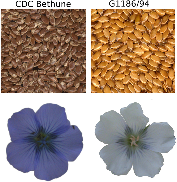
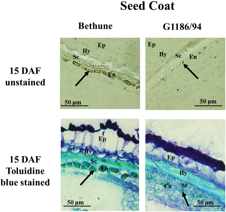
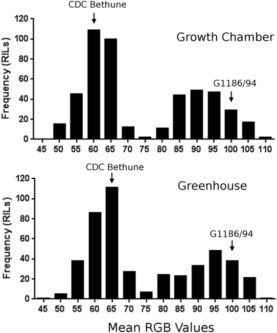
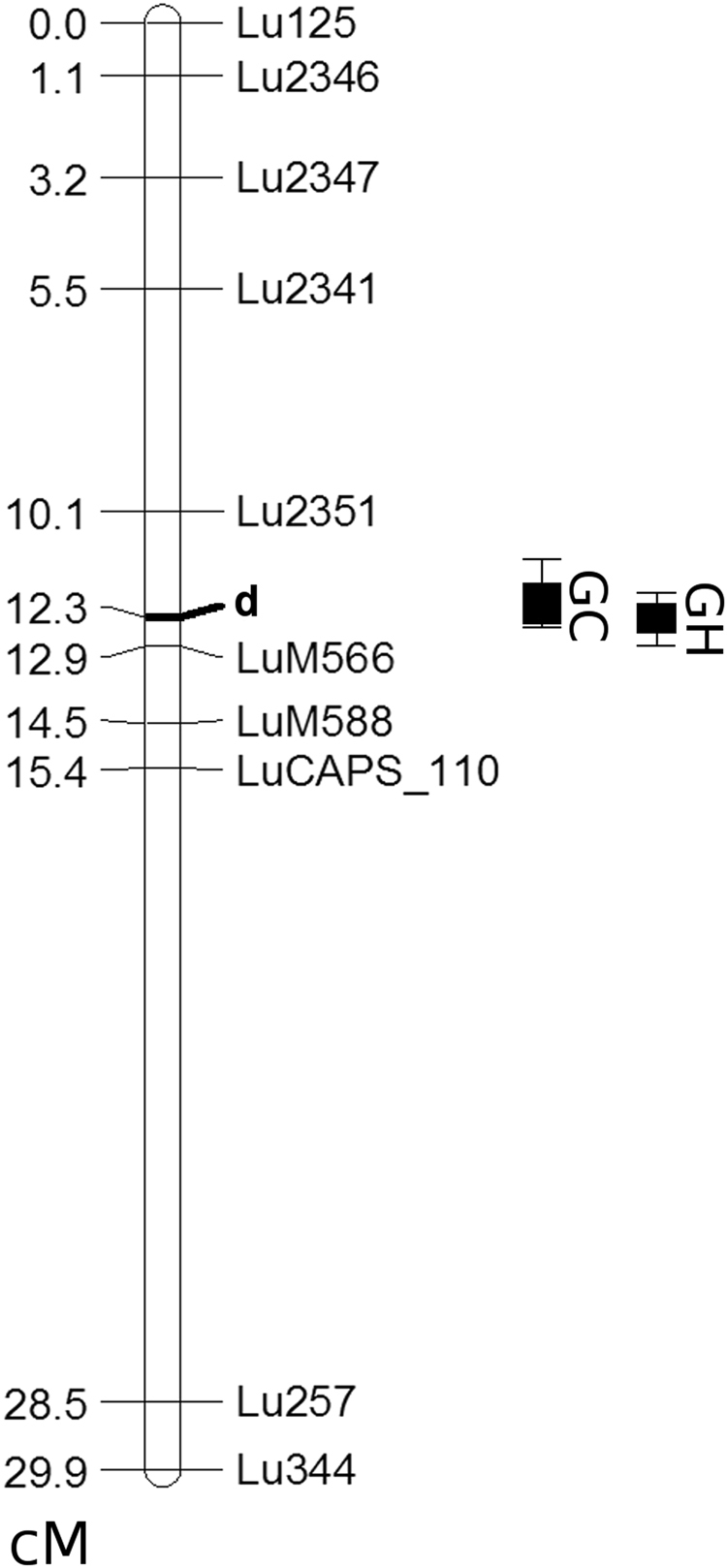
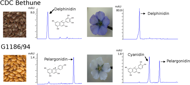
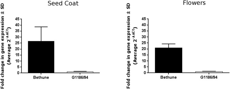
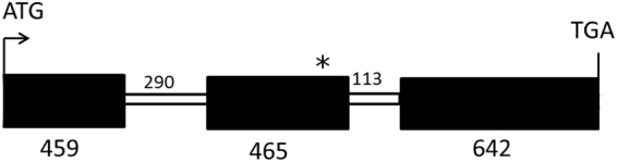
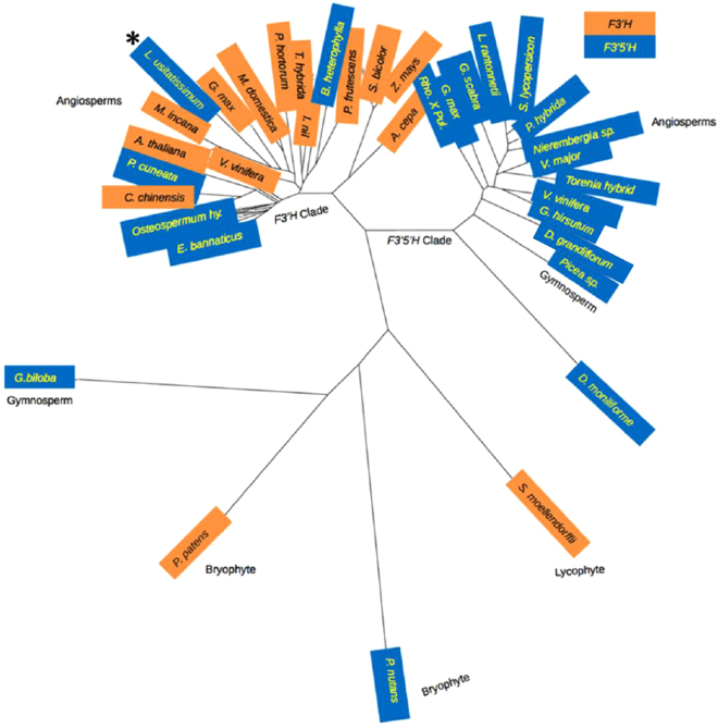
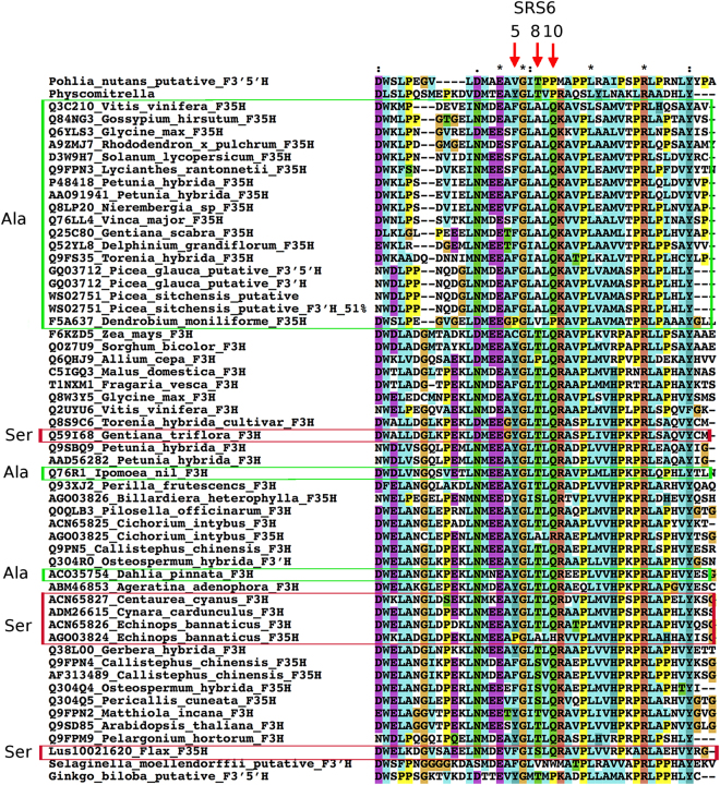
The flowers of flax (linseed) are blue-hued, ephemeral and self-pollinating, and the seeds are typically brown. A century-old interest in natural yellow seed variants and a historical model point to recessive alleles in B1, D and G loci being responsible, but the functional aspects had remained unknown. Here, we characterized the "D" locus by quantitative trait loci (QTL) mapping and identified a FLAVONOID 3'5' HYDROXYLASE (F3'5'H) gene therein. It does not belong to the F3'5'H clade, but resembles biochemically characterized F3'Hs (flavonoid 3' hydroxylase) but without F3'H activity. The genome lacks other F3'H or F3'H-like genes. The apparent neo-functionalization from F3'H is associated with a Thr498 → Ser498 substitution in a substrate recognition site (SRS). The yellow seed and white flower phenotypes of the classical d mutation was found to be due to one nucleotide deletion that would truncate the deduced product and remove three of the six potential SRS, negatively impacting delphinidin synthesis. Delphinidin is sporadic in angiosperms, and flax has no known pollination syndrome(s) with functional pollinator group(s) that are attracted to blue flowers, raising questions on the acquisition of F3'5'H. The appearance of d allele is suggestive of the beginning of the loss of F3'5'H in this species.


Conflict of interest statement

The authors declare that they have no competing interests.

Figures

Figure 1
Figure 1
Seed and flower color phenotypes of CDC Bethune (a representative of the wild type; brown seed and blue-hued petals) and G1186/94 (d mutant with yellow seed and white petals).
Figure 2
Figure 2
Deficiency in pigment accumulation in the seed coat of G1186/94 (yellow seed) in contrast to CDC Bethune (brown seeds) as shown in cross sections of seeds at 15 days after flowering (DAF). The two upper panels are unstained sections, and the two bottom panels are sections stained with toluidine blue. The dark coloration in both the unstained and stained CDC Bethune endothelium is indicative of the presence of proanthocyanidins (PA) as indicated by the arrows. The corresponding cell layer in G1186/94, which is deficient in PA accumulation, is also indicated by arrows. (Ep- epidermis, Hy- hypodermis, Sc-sclerenchyma layer, En- endothelium).
Figure 3
Figure 3
Phenotypic distribution of seed color in G1186/94 X CDC Bethune recombinant inbred line (RIL) population grown in two different environments (a controlled growth cabinet and a less controlled greenhouse). Brown seeds (e.g. CDC Bethune) had a lower mean RGB value of <70 whereas yellow seeds (e.g. G1186/94) had a value of >80. The parental lines indicated by arrows had the phenotypes shown in Fig. 1.Transgressive segregants for both darker brown and brighter yellow were evident in both environments.
Figure 4
Figure 4
Fine mapping of the D locus in LG2. Recessive mutation d imparts both yellow seed and white petal color. QTL location between Lu2351 and LuM566 in recombinant inbred line population of G1186/94 x CDC Bethune grown in two environments is shown: GC, controlled growth cabinet (LOD 47.1 and R2 0.89); GH, greenhouse (LOD 39.1 and R2 0.84). The genetic location of some markers are not shown in the figure to avoid crowding the depiction, but it is indicated in parentheses, preceding the physical location corresponding to the nucleotide sequence of Scaffold 208: LuM566 (12.9), 53.5 kb; LuM568 (12.9), 58.5 kb; LuM569 (12.9), 58.6 kb;. LuM588 (14.5), 141.2 kb; LuM592 (14.5), 152.5 kb; LuM193 (14.5), 155.1 kb; LuM595 (14.5), 155.6 kb; LuM597 (14.5), 159.9 kb; Lu209 (14.5), 184.1 kb; LuM71 (15.4), 423.5 kb; LuCAPS_110, 447.7 kb. The primer information for the LuM SSR markers is provided in Supp. Table S1. LuCAPS_110 is a CAPS marker that is described in Methods. (cM = centimorgans).
Figure 5
Figure 5
Qualitative and quantitative differences in the flavonoids from seeds and petals of CD Bethune and G1186/94 as shown by HPLC profiles of acidified butanol extracts. Note the compressed scale for CDC Bethune and expanded scale for G1186/94. CDC Bethune flowers also contained a similar amount of cyanidin as in G1186/94 flowers.
Figure 6
Figure 6
Lower expression of F3′5′H gene in G1186/94 as identified from qRT-PCR analysis of transcripts in seed coat (15 days after flowering) and petals (flower buds at anthesis before opening). EF1α was used as the common internal control reference gene. Higher level of expression in CDC Bethune relative to G1186/94 is shown.
Figure 7
Figure 7
Depiction of F35H gene in CDC Bethune and G1186/94. Exons are shown as filled boxes and introns as open boxes. The number of nucleotides in each of these is as indicated. The asterisk denotes a premature stop codon created due to a single nucleotide deletion in G1186/94. Further details are in Supplementary Figures 3 and 4. The sequence between ATG and TGA shown in the figure was amplified with suitable primers highlighted in Supplementary Figure 3. An alignment of CDC Bethune and G1186/94 amplicon sequences is shown in Supplementary Figure 4.
Figure 8
Figure 8
Location of the flax F3′5 H (marked with an asterisk) in the F3′H clade. The unrooted phylogenetic tree was constructed as described in Methods. The accessions are from BRENDA (http://www.brenda-enzymes.org), Seitz et al., and NCBI. The species names and the entries are as in Fig. 9. The enzymatic function is known for all entries except those from Ginkgo biloba, Picea glauca, Physcomitrella patens, Pohlia nutans, Selaginella moellendorffii.
Figure 9
Figure 9
Alignment of putative F3′H and F3′5′H amino acid sequences with marked region for substrate recognition site (SRS6). The accessions are from BRENDA (http://www.brenda-enzymes.org), Seitz et al., and NCBI. The positions 5, 8 and 10 within SRS6 are indicated by arrows. Notable for substrate specificity is position 8,. The accessions where a Ser (S) is present at this site instead of Thr (T) are boxed in red; Ala (A) substituted sequences are boxed in green. These boxed proteins are F3′5′H whereas others are F3′H. “Lus10021620_Flax_F35H” boxed in red is the flax D locus gene product that has a Ser substitution at position 8.

Similar articles

Cited by

References

    1. Allaby RG, Peterson GW, Merriwether DA, Fu YB. Evidence of the domestication history of flax (Linum usitatissimum L.) from genetic diversity of the sad2 locus. Theor Appl Genet. 2005;112:58–65. doi: 10.1007/s00122-005-0103-3. - DOI - PubMed
    1. Kvavadze E, et al. 30,000-year-old wild flax fibers. Science. 2009;325:1359. doi: 10.1126/science.1175404. - DOI - PubMed
    1. Green, A. G. & Dribnenki, J. C. P. Breeding and development of LINOLA (low-linolenic flax). In The Third Meeting of the International Flax Breeding Group. 144–150 (1995).
    1. Shirley BW, et al. Analysis of Arabidopsis mutants deficient in flavonoid biosynthesis. The Plant Journal. 1995;8:659–671. doi: 10.1046/j.1365-313X.1995.08050659.x. - DOI - PubMed
    1. Xu W, Dubos C, Lepiniec L. Transcriptional control of flavonoid biosynthesis by MYB–bHLH–WDR complexes. Trends in Plant Science. 2015;20:176–185. doi: 10.1016/j.tplants.2014.12.001. - DOI - PubMed

LinkOut - more resources