. 2017 Nov 16;7(1):15714.
doi: 10.1038/s41598-017-15953-x.

Revisiting the role of interleukin-8 in chronic lymphocytic leukemia

Affiliations

Revisiting the role of interleukin-8 in chronic lymphocytic leukemia

Denise Risnik et al. Sci Rep. .

Abstract

The proliferation and survival of malignant B cells in chronic lymphocytic leukemia (CLL) depend on signals from the microenvironment in lymphoid tissues. Among a plethora of soluble factors, IL-8 has been considered one of the most relevant to support CLL B cell progression in an autocrine fashion, even though the expression of IL-8 receptors, CXCR1 and CXCR2, on leukemic B cells has not been reported. Here we show that circulating CLL B cells neither express CXCR1 or CXCR2 nor they respond to exogenous IL-8 when cultured in vitro alone or in the presence of monocytes/nurse-like cells. By intracellular staining and ELISA we show that highly purified CLL B cells do not produce IL-8 spontaneously or upon activation through the B cell receptor. By contrast, we found that a minor proportion (<0.5%) of contaminating monocytes in enriched suspensions of leukemic cells might be the actual source of IL-8 due to their strong capacity to release this cytokine. Altogether our results indicate that CLL B cells are not able to secrete or respond to IL-8 and highlight the importance of methodological details in in vitro experiments.


Conflict of interest statement

The authors declare that they have no competing interests.

Figures
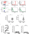
Figure 1
Figure 1
CLL cells do not express IL-8 receptors CXCR1 or CXCR2. Neutrophils (R1) and PBMC (R2) were first discriminated by size (FSC-H) and internal complexity (SSC-H). Leukemic cells were further identified by CD19 expression (R3). (a) CXCR1 and CXCR2 expression in CLL cells and neutrophils evaluated by flow cytometry. Shown are representative histograms from one sample with isotype control for each IL-8 receptor in grey line. (b) Graphs show mean fluorescence intensity (MFI) of CXCR1 and CXCR2 in neutrophils and CLL cells (mean ± SEM n = 56). Statistical analysis was performed using Mann-Whitney test. Asterisks indicate statistically significant differences (***p < 0.001). Activation of CLL cells does not induce the expression of IL-8 receptors. PBMC (3 × 106/ml) were incubated with immobilized anti-IgM plus CD40L (40 ng/ml) or medium alone (control) for 24 h. Shown are the percentages of CD69+ (c), CXCR1+ and CXCR2+ (d) leukemic B cells under control or activated conditions (n = 10). Statistical analysis was performed using Wilcoxon test. *p < 0.05, ns: not significant).
Figure 2
Figure 2
IL-8 does not prolong CLL cell survival in vitro. PBMC or monocyte-depleted PBMC (3 × 106/ml) were incubated with or without recombinant IL-8 (20 ng/ml) for 10 days. IL-8 was added on days 0, 3, 6 and 9. Cell death of CD19+ lymphocytes was evaluated by flow cytometric alterations of light-scattering properties and confirmed by Annexin V staining on days 4, 7 and 10. Shown are mean ± SEM, n = 9. Statistical analysis was performed using Friedman test followed by the Dunn´s multiple comparison post-test. Asterisks indicate statistically significant differences (***p < 0.001) between PBMC and monocyte-depleted PBMC. The addition of IL-8 did not significantly modify cell survival of PBMC or monocyte-depleted PBMC.
Figure 3
Figure 3
Monocytes, but not leukemic B cells, produce IL-8 in PBMC samples from CLL patients. (a) PBMC (3 × 106/ml) were incubated with immobilized anti-IgM plus CD40L (40 ng/ml) or medium alone (control) for 24 h. Monensin (20 μM) was added for the last 4 h. to inhibit Golgi transport. IL-8 expression was evaluated by intracellular staining in CD19+ and CD14+ cells. Shown are representative dot plots from one CLL sample (left) and the percentages of IL-8 positive cells (mean ± SEM, n = 17) (right). (b) Aliquots of PBMC from 13 CLL samples were depleted from monocytes as described in Methods. The percentage of monocytes in each sample before and after depletion is shown. (c) Different cell numbers of PBMC or the corresponding monocyte-depleted PBMC were incubated with immobilized anti-IgM plus CD40L (40 ng/ml) or medium alone (control) for 24 h. The levels of IL-8 released to supernatants were quantified by ELISA. Results show the mean ± SEM of the 13 CLL samples. Statistical analysis was performed using Friedman test followed by the Dunn post-test. Asterisks indicate statistically significant differences between PBMC and monocyte-depleted PBMC (*p < 0.05; ***p < 0.001), both under control or activated conditions.

References

    1. Chiorazzi N, Rai KR, Ferrarini M. Chronic lymphocytic leukemia. The New England journal of medicine. 2005;352:804–815. doi: 10.1056/NEJMra041720. - DOI - PubMed
    1. Kipps TJ, et al. Chronic lymphocytic leukaemia. Nature reviews. Disease primers. 2017;3:17008. doi: 10.1038/nrdp.2017.8. - DOI - PubMed
    1. Burger JA, Gribben JG. The microenvironment in chronic lymphocytic leukemia (CLL) and other B cell malignancies: insight into disease biology and new targeted therapies. Seminars in cancer biology. 2014;24:71–81. doi: 10.1016/j.semcancer.2013.08.011. - DOI - PubMed
    1. di Celle PF, et al. Cytokine gene expression in B-cell chronic lymphocytic leukemia: evidence of constitutive interleukin-8 (IL-8) mRNA expression and secretion of biologically active IL-8 protein. Blood. 1994;84:220–228. - PubMed
    1. Wierda WG, et al. Plasma interleukin 8 level predicts for survival in chronic lymphocytic leukaemia. British journal of haematology. 2003;120:452–456. doi: 10.1046/j.1365-2141.2003.04118.x. - DOI - PubMed

LinkOut - more resources