Case Reports
. 2014 Apr;5(2):90-92.
doi: 10.14740/wjon783w. Epub 2014 May 6.

Adrenal Ganglioneuroma Presenting As Left Renal Mass

Affiliations
Case Reports

Adrenal Ganglioneuroma Presenting As Left Renal Mass

Hakan Ozturk. World J Oncol. 2014 Apr.

Abstract

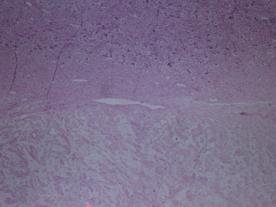
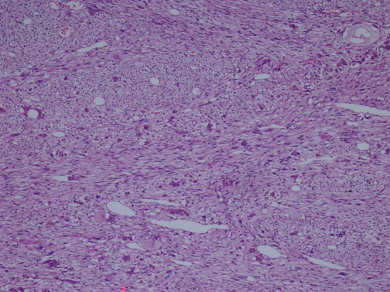
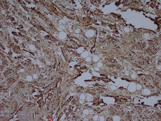
Ganglioneuromas (GNs) are benign tumors resulting from neural crest tissue. GNs contain mature ganglion cells and Schwann cells. GNs most commonly occur in the retroperitoneum and posterior mediastinum. GNs rarely occur in the adrenal gland. A 45-year-old asymptomatic patient presented with an incidental finding of left renal mass. A 10 cm mass lesion located in the upper pole of the left kidney and lymphadenopathy in renal hilus were detected. The patient underwent transperitoneal radical nephrectomy involving the removal of left adrenal gland. The immunohistochemical examination showed strong positive staining for S100, neuron-specific enolase, synaptophysin and chromogranin. The diagnosis of mature GN was established. GNs are among the rare diseases that should be considered in the evaluation of renal masses, particularly in the differential diagnosis of upper pole tumors of the kidneys. It can be confused with renal cell carcinomas.

Keywords: Adrenal ganglioneuroma; Ganglioneuroma; Immunohistochemistry; Incidentaloma; Renal mass.


Figures

Figure 1
Figure 1
Hematoxylin-eosin staining demostrating H&E (× 40).
Figure 2
Figure 2
Hematoxylin-eosin staining demostrating H&E (× 100).
Figure 3
Figure 3
Immunohistochemistry showing S100 (× 100).

Similar articles

References

    1. Anagnostis P, Karagiannis A, Tziomalos K, Kakafika AI, Athyros VG, Mikhailidis DP. Adrenal incidentaloma: a diagnostic challenge. Hormones (Athens) 2009;8(3):163–184. doi: 10.14310/horm.2002.1233. - DOI - PubMed
    1. Geoerger B, Hero B, Harms D, Grebe J, Scheidhauer K, Berthold F. Metabolic activity and clinical features of primary ganglioneuromas. Cancer. 2001;91(10):1905–1913. doi: 10.1002/1097-0142(20010515)91:10<1905::AID-CNCR1213>3.0.CO;2-4. - DOI - PubMed
    1. Ugarriza LF, Cabezudo JM, Ramirez JM, Lorenzana LM, Porras LF. Bilateral and symmetric C1-C2 dumbbell ganglioneuromas producing severe spinal cord compression. Surg Neurol. 2001;55(4):228–231. doi: 10.1016/S0090-3019(01)00363-9. - DOI - PubMed
    1. Lonergan GJ, Schwab CM, Suarez ES, Carlson CL. Neuroblastoma, ganglioneuroblastoma, and ganglioneuroma: radiologic-pathologic correlation. Radiographics. 2002;22(4):911–934. doi: 10.1148/radiographics.22.4.g02jl15911. - DOI - PubMed
    1. Ma J, Liang L, Liu H. Multiple cervical ganglioneuroma: A case report and review of the literature. Oncol Lett. 2012;4(3):509–512. - PMC - PubMed

Publication types

LinkOut - more resources